

"I have used a wide variety of secondaries and Jackson ImmunoResearch has consistently been the best. The fluorophores are bright and stable and their selective (x reactivity removed) secondaries have always shown species specificity in multiple labeling."
Janet Duerr, Ohio UniversityRating: 5.0
The following information details how to use the Product Filter on the left of the page to select secondary antibodies and reagents. It may also be helpful when using the product tables in our catalog and online.
See also our guidance on generating product descriptions.
Affinity-purified secondary antibodies are offered in three different formats, and the optimal choice depends on the intended application. Select from Whole IgG, F(ab')2 fragment, or Fab fragment antibodies.
The whole IgG form of antibodies is suitable for the majority of immunodetection procedures. F(ab')2 and Fab antibodies may be indicated for experiments with specific assay requirements.
Antisera have not been affinity-purified.
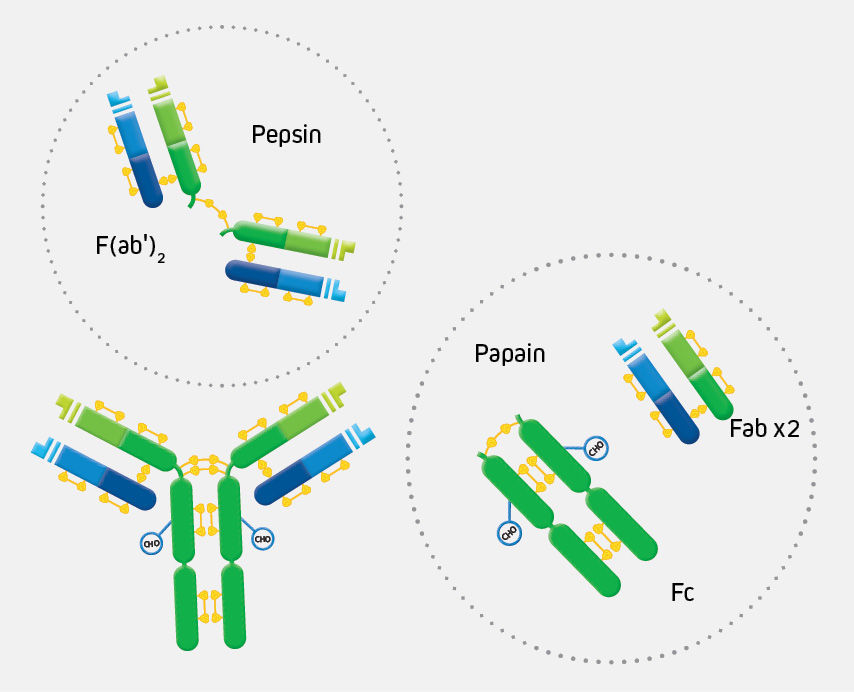
Whole IgG antibodies are isolated from antisera by immunoaffinity chromatography. They have an Fc region and two antigen binding Fab regions joined together by disulfide bonds (Figure 1), and therefore they are divalent. The average molecular weight is reported to be about 160 kDa. The whole IgG form of antibodies is suitable for the majority of immunodetection procedures and is the most cost effective.
Read more about whole IgG format secondary antibodies.F(ab')2 fragment antibodies are generated by pepsin digestion of whole IgG antibodies to remove most of the Fc region while leaving intact some of the hinge region. F(ab')2 fragments have two antigen-binding Fab regions linked together by disulfide bonds, and therefore they are divalent. The average molecular weight is about 110 kDa. They are used for specific applications, such as to avoid binding of secondary antibodies to live cells with Fc receptors or to Protein A or Protein G.
Read more about F(ab')2 fragment secondary antibodiesBinding of primary antibodies to Fc receptors also may occur if they are whole IgG antibodies, creating background regardless of the form of the secondary antibody. To block whole IgG primary and secondary antibodies from binding to Fc receptors, incubate cells in buffer containing 5% normal serum from the host species of the labeled secondary antibody.
To prevent capping, endocytosis, and regeneration of Fc receptors on living cells, incubate at 4°C in buffer containing 5% normal serum with sodium azide added to inhibit metabolism. More information on avoiding background.
Fab fragment antibodies are generated by papain digestion of whole IgG antibodies to remove the entire Fc portion, including the hinge region (Figure 1). These antibodies are monovalent, containing only a single antigen binding site. The molecular weight of Fab fragments is about 50 kDa. They can be used to block endogenous immunoglobulins on cells, tissues or other surfaces, and to block the exposed immunoglobulins in multiple labeling experiments using primary antibodies from the same species.
Read more about Fab fragment secondary antibodies.FabuLight™ antibodies are Fab fragment secondary antibodies specific to the Fc region of IgG or IgM primary antibodies. They enable labeling of primary antibodies prior to incubation with cells or tissue without compromising the active site of the primary antibody. They can also be used to label cell surface immunoglobulins without cross-linking and activating B cells.
Read more about FabuLight™.Antisera are commonly used for immunoprecipitation assays such as immunodiffusion and immunoelectrophoresis, but are not suitable for coating ELISA plates or other applications requiring affinity-purified antibodies. Antisera against whole IgG molecules (Anti-IgG (H+L)) are recommended for bridging PAP to primary antibodies.
Read more about antisera.Antibodies are listed alphabetically according to the host species of the primary antibody. For example, if the primary antibody is made in mouse, an “Anti-Mouse” secondary antibody is required.
Note: Both anti-Syrian and anti-Armenian hamster secondary antibodies are listed under “Anti-Hamster”. It is important to know in which strain of hamster the primary antibody was produced since cross-reaction between the strains is not complete.
Selection of the host species for a secondary antibody involves many considerations, including but not limited to:
The following explanations of terms may assist in selecting the most appropriate antibody specificity for the experiment.
Note: Immunoglobulins from different species share similar structures, with similarities being related to closeness in phylogeny. Antibodies against immunoglobulins from one species are likely to cross-react with a number of other species, unless they have been specifically adsorbed against the cross-reacting species. Antibodies that have been adsorbed against other species will contain “(min X...Sr Prot )” in the antibody description (see step 5).
These antibodies react with both the heavy and light chains of the IgG molecule, i.e. with both the Fc and F(ab')2 / Fab regions of IgG (Figure 1). Anti-IgG (H+L) antibodies also react with other immunoglobulin classes (IgM, IgA, IgD, IgE) and subclasses since they all share the same light chains (either kappa or lambda). Anti-IgG (H+L) antibodies have broader epitope recognition than anti-fragment specific antibodies. They are suggested for all general immunodetection procedures.
These antibodies react with the light chains shared by IgG and the other immunoglobulins. They were developed to facilitate detection of proteins around 50 kDa on Western blots after immunoprecipitation (IP), and do not react with IgG heavy chains.
Read more about Western blotting after Immunoprecipitation.These antibodies react with the F(ab')2 / Fab portion of IgG. They have been tested by ELISA and/or adsorbed against Fc fragments. They are not specific for IgG since they react with light chains, and therefore also react with other immunoglobulin classes (IgA, IgM, IgD, and IgE) and subclasses sharing the same light chains.
These antibodies react with the VHH domain of alpaca and llama IgG subclasses 2 and 3. Read more about secondary antibodies for VHH discovery.
These antibodies react with the Fc portion of the IgG heavy chain. They have been tested by ELISA and/or adsorbed against Fab fragments. In some cases, they are additionally tested and/or adsorbed to minimize cross-reactivity to IgM and/or IgA. In such cases (anti-human, anti-mouse, and anti-rat), they are labeled “Anti-IgG, Fcγ”.
Caution: Anti-IgG, Fcγ fragment specific antibodies do not react equally with all monoclonal primary antibodies. For an anti-mouse IgG, Fcγ fragment specific antibody with balanced reactivity to four subclasses of IgG, select Goat Anti-Mouse IgG (subclasses 1+2a+2b+3), Fcγ fragment specific (min X Hu, Bov, Rb Sr Prot).
These antibodies react with the Fc portion of the heavy chain of individual subclasses of mouse IgG. They have been tested by ELISA and/or adsorbed to minimize cross-reactivity to other subclasses, Fab fragments and IgM; and human, bovine and rabbit serum proteins. Anti-Mouse IgG, Fcγ subclass specific antibodies react with individual subclasses of mouse IgG. They are intended for distinguishing between different subclasses of mouse IgG primary antibodies in multiple labeling experiments, or for IgG subclass determination
Read more about Anti-Mouse IgG, Fcγ subclass specific antibodies.These antibodies react with heavy chains of IgG and IgM. They also react with the light chains that are shared among immunoglobulins, so they may also react with IgA, IgD and IgE.
These antibodies react with heavy chains of human IgA, IgG and IgM. They also react with the light chains that are shared among human immunoglobulins, so they may also react with IgD and IgE.
These antibodies react with the heavy chain of IgM (in the case of anti-human, just the Fc5μ portion of the heavy chain). They have been tested by ELISA and/or adsorbed against IgG. Anti-human IgM, Fc5μ is additionally tested and/or adsorbed to minimize cross-reactivity to IgA.
These antibodies react with the heavy chain of human IgA. They have been tested by ELISA and/or adsorbed to minimize cross-reactivity with human IgG and IgM.
Jackson ImmunoResearch Anti-Human IgE antibodies are available in two different clones (ME.114 and 10A10) and are both mouse-derived monoclonal antibodies with reactivity to human E class immunoglobulins and are epsilon (ε) chain specific.
Anti-Fluorescein, Anti-HRP, Anti-Biotin and Anti-Digoxin are available for labeling endogenous proteins or nucleic acid probes and for signal enhancement or signal conversion.
Read more about signal enhancement and conversion.Secondary antibodies against one species are likely to cross-react with other species unless they have been specifically adsorbed against the other species. Antibodies with “(min X ... Sr Prot)” in the description have been tested and/or adsorbed against IgG and/or serum proteins of those species indicated in the parentheses. They are recommended when the presence of immunoglobulins from other species may lead to interfering cross-reactivities.
Read more about cross-adsorbed secondary antibodies.| min X = minimal cross-reaction | Ar Hms = Armenian Hamster | Rb = Rabbit |
| Bov = Bovine | Sy Hms = Syrian Hamster | Shp = Sheep |
| Ck = Chicken | Hrs = Horse | Sw = Swine |
| Gt = Goat | Hu = Human | Sr = Serum |
| GP = Guinea Pig | Ms = Mouse | Prot = Protein |
Note: Caution should be exercised when considering antibodies that have been adsorbed against closely related species, since they have greatly reduced epitope recognition and may recognize some monoclonals poorly. For example, choose anti-mouse IgG adsorbed against rat IgG to detect a mouse primary antibody in rat tissue which contains endogenous rat immunoglobulins, or in a multiple labeling application which includes a rat primary antibody. Use anti-mouse IgG not adsorbed against rat IgG to detect a mouse primary antibody in the absence of rat immunoglobulins. Two other examples of antibodies which have diminished epitope recognition after adsorption with closely related species are Anti-Rat IgG (Min X ... Mouse ... Sr Prot) and Anti-Armenian Hamster IgG (min X ... Mouse, Rat ... Sr Prot).
Some antibodies are designated ML to emphasize their usefulness in multiple labeling in addition to single labeling. Read more about multiple labeling with secondary antibodies.
Most hamster monoclonal antibodies are derived from Armenian hamster spleen cell-mouse myeloma hybridomas. The IgG produced by these hybridomas is Armenian hamster IgG, while most polyclonal hamster antibodies are raised in Syrian hamsters. Antibodies raised against one hamster species are not as effective in detecting the other species, so it is important to know the origin of a hamster primary antibody.
Caution: Anti-Armenian Hamster IgG (H+L) (min X Bov, Hu, Ms, Rb, Rat Sr Prot) may not recognize all Armenian hamster monoclonal antibodies, since it has been adsorbed against closely related species (in bold). Therefore, it is better to use an antibody adsorbed against fewer species, such as Anti-Armenian Hamster IgG (H+L) (min X Bov Sr Prot), except in those cases where Armenian hamster monoclonals need to be detected in the presence of mouse and/or rat immunoglobulins.
In addition to unconjugated antibodies, JIR offers antibodies conjugated to a wide range of probes. Fluorescent dyes, fluorescent proteins, reporter enzymes, Biotin-SP™ and colloidal gold are among the conjugate choices.
Read more about conjugate selection.Depending on the technique, other immunoreagents may be required to optimize the assay. Blocking reagents, experimental controls and signal enhancement molecules are available in a variety of formats.
Read more about blocking, controls and diluents.Streptavidin reagents are available for use with biotinylated antibodies.
