

"I have used a wide variety of secondaries and Jackson ImmunoResearch has consistently been the best. The fluorophores are bright and stable and their selective (x reactivity removed) secondaries have always shown species specificity in multiple labeling."
Janet Duerr, Ohio UniversityRating: 5.0
Our DyLight 405-conjugated secondary antibodies are excited maximally at about 400 nm and fluoresce with a peak at about 421 nm. They are very bright and photostable when used with microscopes or flow cytometers with a 405 nm laser and a 420 nm emission filter (Figure 2). They will not work well with a 450 nm emission filter.
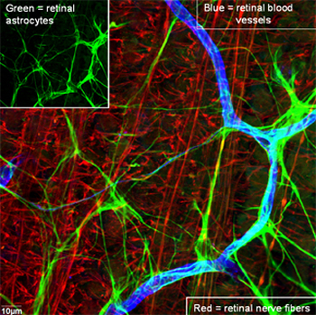
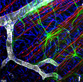
Under these conditions, it is possible to perform effective 4-color imaging with good color separation, good photostability, and high sensitivity in both aqueous and permanent mounting media. The combination of DyLight 405, Alexa Fluor 488, Rhodamine Red-X, and Alexa Fluor 647 provides for maximum color separation (see Spectra Viewer).
Other 4-color dye combinations, which may be equally effective but have slightly less color separation, include DyLight 405, Alexa Fluor 488, Cy3, and Alexa Fluor 647.
The following table lists highly adsorbed affinity-purified secondary antibodies, streptavidin, and immunoglobulin controls conjugated with DyLight 405 as offered by Jackson ImmunoResearch for use only in 4-color labeling protocols using a 405 nm laser-equipped confocal microscope. For a complete listing of other DyLight 405 conjugates for 1-, 2-, and 3-color labeling, see complete tables of Whole IgG Secondary Antibodies and F(ab')2 Fragment Secondary Antibodies.
| Product Description | Product Code |
|---|---|
| Streptavidin | 016-470-084 |
| ChromPure Donkey IgG, whole molecule | 017-470-003 |
| ChromPure Goat IgG, whole molecule | 005-470-003 |
