
The secondary antibody detects the primary antibody, typically conjugated to a reporter molecule it enables the visualization of the protein(s) of interest. Part 6 of our Western blotting guide details hints, tips and best practices for handling your secondary antibodies for Western blotting.
Overview
Following incubation with the primary antibody and thorough washing, the membrane is incubated with the secondary antibody. The secondary antibody binds the primary antibody and is typically conjugated to a reporter molecule which enables the visualization of the protein(s) of interest.
Advantages of Indirect Detection
Western blotting is typically performed indirectly, using a conjugated secondary antibody instead of a directly conjugated primary antibody. Indirect detection affords greater assay sensitivity and reagent flexibility. Multiple secondary antibodies can bind each primary antibody, and each secondary antibody can bring multiple reporter molecules to the antibody complex, amplifying the signal from each primary antibody.
Indirect detection preserves the affinity of the primary antibody by eliminating the possibility of conjugation interfering with its antigen-binding sites (paratopes), preventing compromised detection of the target molecule antigens.
Directly conjugated primary antibodies are often of limited availability, with a poor range of species or conjugate offered commercially. Greater choice is possible using indirect detection. Secondary antibodies are readily available conjugated to a wide range of reporter molecules, from enzymes, fluorescent dyes and proteins, and to amplification molecules such as biotin.
Indirect detection also enables one secondary antibody to be used with a variety of primary antibodies, which may present better value than purchasing multiple direct conjugates.
Considerations for Choosing a Secondary Antibody – Host Species
Compatibility of Host Species
If using multiple secondary antibodies, for example probing for two primary antibodies, choose the same host species to minimize any cross-reactivity. If this is not possible, then choose secondary antibodies that are cross-adsorbed against the host species of the other secondary antibodies in the experiment.
Cross-Reacting Species
Although not common in western blot, some applications require secondary antibodies that are adsorbed against other species to minimize recognition of endogenous immunoglobulins (such as probing mouse tissue lysate with an anti-rat primary antibody)
or other primary antibodies. Therefore, choose a secondary antibody host that is available with the appropriate cross-adsorptions for the intended application.
Experience or Preference
The choice of host species often comes down to either personal preference or previous experimental validation. There appears to be little difference in quality between secondary antibodies raised in different host species, merely that some species are more appropriate for large-scale production.
Specificity
It is important to pick a secondary antibody with the correct specificity. Although whole IgG (H+L) is suitable for most immunoassays, other specialist specificities allow more complicated experiments to be designed to remove potential interference from unrequired detection.
Anti-IgG (H+L)
These antibodies react with both the heavy and light chains of the IgG molecule, i.e., with both the Fc and F(ab’)2 / Fab regions of IgG (Figure 1). Anti-IgG (H+L) antibodies also react with other immunoglobulin classes (IgM, IgA, IgD, IgE) and subclasses since they all share the same light chains (either kappa or lambda).
Anti-IgG (H+L) antibodies have broader epitope recognition than anti-fragment specific antibodies. They are also suggested for all general immunodetection procedures.
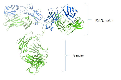
Figure 1: Ribbon diagram of IgG and F(ab’)2 and Fc region
Isotype Specificity: Fc Specific Antibodies
Most primary antibodies are of the IgG isotype. Other classes, such as IgM and IgA, are sometimes used depending on how the antibody has been generated. Secondary antibodies which are specific to the Fc domain of the immunoglobulin will be specific to the isotype of that particular Fc domain.
Therefore, an Fcγ specific antibody will be specific to IgG, whereas an antibody specific to Fc α will detect IgA specifically, and Fcμ will detect IgM.
Pan-specific antibodies are also available, which can be used to detect two or more isotypes. Depending on your application, this may be useful. Choose a secondary antibody that is specific to the isotype of the primary antibody.
Subclass Specificity
IgG-specific antibodies will detect all gamma isotype immunoglobulins due to the similarity of their homologous Fcγ domains. Depending on the mouse strain used to generate the primary antibody, the subclass of the immunoglobulin could be 1 of 5. It may be the case that you have two mouse antibodies of different subclasses targeting two proteins in the same experiment. To differentiate between the two primary antibodies, you would require subclass-specific antibodies.
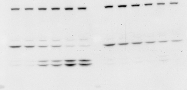
Mice express four of the five available IgG subclasses making up their IgG isotype. They typically produce IgG1, IgG2b and IgG3, and depending on their strain, they will also express either IgG2a or IgG2c.¹ Jackson ImmunoResearch Anti-Mouse IgG subclass specific antibodies offer specificity to the five individual mouse IgG subclasses. These highly specific antibodies are designed to distinguish between two or more different subclasses of mouse IgG in multiplex experiments or for mouse IgG subclass determination. They have been adsorbed against human, bovine and rabbit serum proteins to minimize cross-reactivity with tissue immunoglobulins, adherent bovine IgG on cultured cells, and rabbit primary antibodies. Subclass-specific antibodies provide exquisite discrimination between the subclasses.
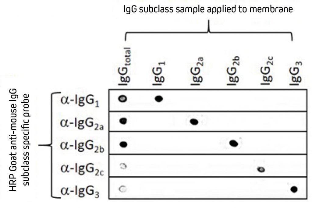 Separate nitrocellulose strips (rows) received 100 ng “dots’’ of mouse IgG and each subclass and then were blocked with 5% (w/v) BSA in PBST. After probing with HRP-conjugated Goat Anti-Mouse subclass-specific antibodies, the strips were developed with TMBM substrate. The grid of positive signals shows the specificity of each subclass-directed antibody. Some subclasses are poorly represented in a total IgG pool and thus give a weak signal (IgG total vs. Anti-IgG2c and -IgG3). HRP-conjugates used for probing were 115-035-205 (Anti-Mouse IgG1), 115-035-206 (Anti-Mouse IgG2a), 115-035-207 (Anti-Mouse IgG2b), 115-035-208 (Anti-Mouse IgG2c) and 115-035-209 (Anti-Mouse IgG3). |
CautionAnti-IgG, Fcγ fragment specific antibodies do not react equally with all monoclonal primary antibodies. For an anti-mouse IgG, Fcγ fragment specific antibody with balanced reactivity to four subclasses of IgG, select Goat Anti-Mouse IgG (subclasses 1+2a+2b+3), Fcγ fragment specific (min X Hu, Bov, Rb Sr Prot). Additionally, subclass-specific antibodies are not necessary for general detection of mouse monoclonal antibodies in single-labeling experiments or multiple-labeling experiments involving one mouse monoclonal and primary antibodies from other species. |
Anti-IgG: Light Chain Specific
These antibodies react with the light chains shared by IgG and the other immunoglobulins. They were developed to facilitate the detection of proteins around 50 kDa on western blots after immunoprecipitation (IP) and do not react with IgG heavy chains.
| Western Blotting after Immunoprecipitation
For researchers who perform western blotting following immunoprecipitation, antibodies specific for light chains or Fc fragments allow unobstructed detection of antigens in the 50 kDa or 25 kDa ranges, respectively. Anti-Light Chain Specific AntibodiesWhen labeled secondary antibodies specific for both heavy and light chains of IgG (such as anti-IgG (H+L)) are used to detect protein bands on western blots following immunoprecipitation (IP), two bands appear (see Figure 3) corresponding to the heavy (50 kDa) and light (25 kDa) chains of the precipitated primary antibody. These bands usually obscure the detection of any protein of interest with a molecular weight near 50 kDa or 25 kDa. However, when labeled anti-IgG, Light Chain Specific antibodies are used for detection; they bind only to the light chain band on the blot (Figure 3) and to light chains on the native primary antibodies used for detection. Therefore, a 50 kDa protein may be detected on blots without interference from the heavy chain of the precipitating IgG. Light chain-specific antibodies are available directed against goat, mouse, rabbit, rat and sheep. They have been adsorbed to minimize cross-reactivity with immunoglobulins from many other species, which also may be present on blots. 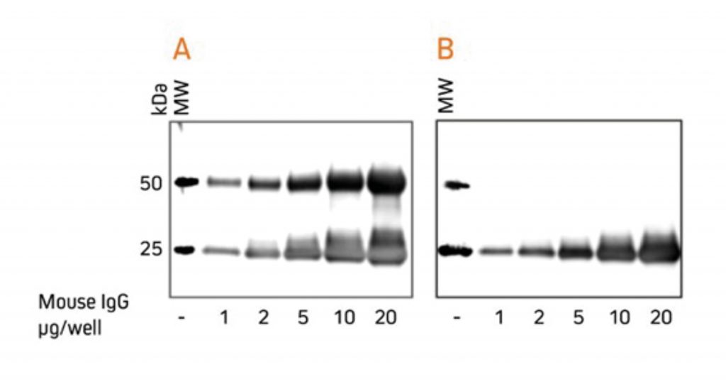 Gels were loaded with reduced and denatured Mouse IgG, whole molecules. After SDS-PAGE and transfer to nitrocellulose, blots were blocked with BSA (10% w/v). After incubation with the secondary antibody, blots were developed with ECL substrate. Blots were imaged simultaneously, with auto exposure time based on bright bands. A: The gel was probed using HRP-conjugated Goat Anti-Mouse IgG (H+L) (115‑035‑003), revealing bands corresponding to both heavy chains (50 kDa) and light chains (25 kDa). B: The gel was probed using HRP-conjugated Goat Anti-Mouse IgG, light chain specific (115‑035‑174), revealing only the 25 kDa band corresponding to Ig light chains. The IP antibody heavy chain is not detected, allowing visualization of the protein of interest near 50 kDa. Heavy Chain Specific Detection on Western Blots after IP with Anti-Fc Specific AntibodiesAnti-IgG, Fc fragment specific antibodies may be used to detect native IgG primary antibodies without binding to the 25 kDa band of reduced and denatured IgG light chains on western blots. Using these antibodies allows clear detection of a 25 kDa analyte without interference from the light chains of an IP antibody. However, this detection is complicated by the appearance of degraded heavy chain antibody at 25 kDa, see Figure 3 panel A. To avoid signal from a degraded heavy chain at 25 kDa, block with monovalent Fab fragment anti-Fc (FabuLight™), see Figure 4, panels B and C. The extreme sensitivity of western blotting requires high concentrations of the blocking reagent. 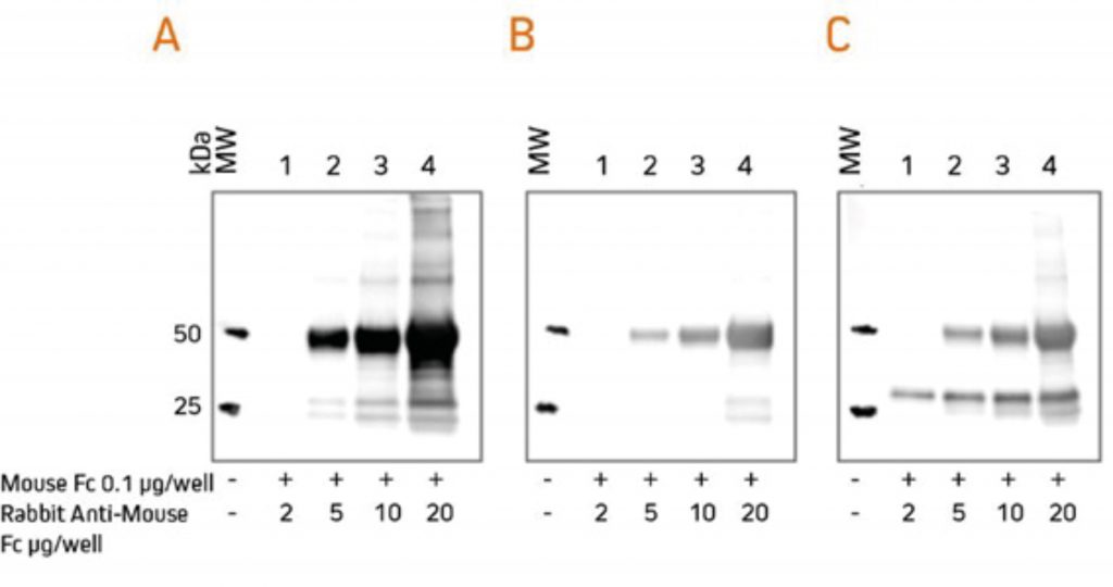 Rabbit Anti-Mouse IgG, Fcγ fragment specific (315-005-008) was mixed with ChromPure™ Mouse Fc (015-000-008) to simulate immunoprecipitation (IP). Three identical gels were run, lanes loaded with denatured and reduced: Lane 1: 2 μg Rabbit Anti-Mouse Fc + 0.1 μg Mouse Fc Lane 2: 5 μg Rabbit Anti-Mouse Fc + 0.1 μg Mouse Fc Lane 3: 10 μg Rabbit Anti-Mouse Fc + 0.1 μg Mouse Fc Lane 4: 20 μg Rabbit Anti-Mouse Fc + 0.1 μg Mouse Fc After SDS-PAGE and transfer to nitrocellulose, blots were blocked with BSA (10% w/v). Subsequent incubations are shown below. After incubation with the secondary antibody, blots were developed with ECL substrate. Blots were imaged simultaneously, with auto exposure time based on bright bands. A: No FabuLight block
The secondary antibody detects the IP antibody heavy chain (HC) at 50 kDa and degraded HC at 25 kDa. B: FabuLight block: Fab Goat Anti-Rabbit IgG, Fc (111-007-008), 200 μg/ml
The signal from IP antibody HC is greatly reduced. At lower loading amounts (2, 5 and 10 μg), the degraded HC at 25 kDa is not detectable. C: FabuLight block: Fab Goat Anti-Rabbit IgG, Fc (111-007-008), 200 μg/ml
The secondary antibody detects the primary antibody, revealing a protein of interest at 25 kDa. Note that the protein of interest shows the sharpest band when the amount of IP antibody is low. To block the signal from a mouse IP antibody, use the Fab fragment corresponding to the subclass of the IP antibody. |
Anti-IgG, F(ab’)2 Fragment Specific
These antibodies react with the F(ab’)2 / Fab portion of IgG. They have been tested by ELISA and/or adsorbed against Fc fragments. They are not specific for IgG since they react with light chains, and they therefore also react with other immunoglobulin classes (IgA, IgM, IgD and IgE) and subclasses sharing the same light chains.
They are predominantly used to detect scFvs (Single-chain variable fragment antibodies). IgG F(ab’)2 fragment specific antibodies may be used in western blotting where enhanced sensitivity to the F(ab’)2 antibody is required and may offer advantages over whole molecule specificity
Using secondary antibodies as a negative control to isolate the cause of non-specific signalA secondary antibody-only control can help to identify the cause of non-specific signals, be that unexpected bands or diffuse signals. Block the membrane with an appropriate blocking solution (5% v/v normal serum of the labeled antibody) and incubate the membrane with the secondary antibody for the prescribed duration, and wash and apply the desired visualization method such as ECL substrate or imager.
|
Determining Optimal Working Concentration
The sensitivity of the secondary antibodies depends on the specificity of the primary antibody and the abundance of the protein of interest. By increasing the concentration of the antibodies, the background signal may increase beyond that of the analyte’s signal.
The correct concentration must be determined to achieve a high signal-to-noise ratio so that background signal does not interfere with detection of the target protein and result interpretation.
Poorly expressed proteins or primary antibodies with weak affinity may benefit from sample concentration or immunoprecipitation rather than extended incubations or more concentrated antibody solutions.
HRP conjugates can offer excellent sensitivity when paired with high-quality chemiluminescent substrates such as ECL, enabling reported antibody dilutions between 1:20,000 and 1:100,000. Optimum working dilution should be determined by titration.
Stepwise dilution of each primary antibody and secondary antibody can be performed easily using a dot blot grid whereby concentrations of antigen and detection reagents can be evaluated and limits of detection determined.
Reprobing and Stripping for Additional Target Proteins
Membranes may be stripped to remove the primary and secondary antibody complex and reprobed with a second pair of antibodies to allow the detection of a second target protein.
The membrane is stripped with a low pH buffer such as 1M glycine to remove the primary and secondary antibody complex from the antigen bound to the membrane. The membrane is then blocked and incubated with a new primary antibody, washed and incubated with a compatible secondary antibody and substrate before being reimaged again.
The success of stripping can be highly variable and must be carefully performed to prevent either being too harsh and damaging the antigen. Alternatively, conditions too mild to strip the antibody complex from the membrane may lead to continued detection.
The results of a reprobed blot should only be used for confirmation, not for quantitation. Choosing a second primary antibody raised in a different host to the initial antibody can help improve the specificity of the reprobing. Stripping and reprobing membranes for additional targets are typically applied in chemiluminescent detection, as fluorescent conjugates offer a more reliable method for detecting multiple targets. See Section 8 for more information about multiplex detection.
|
Caution: Secondary antibody diluent buffer Avoid adding proteins such as powdered milk or BSA to antibody diluent solutions to prevent non-specific interactions that may lead to background signal. Caution: Batch to Batch Variation It is also important to note that the concentration of polyclonal antibodies in serum may vary between batch-to-batch and animal-to-animal, so the dilution curve should be repeated if a change in the analysis is observed. Choose high-quality JIR Affinipure™ affinity-purified antibodies to ensure batch-to-batch consistency between lots. |
| Our rigorous quality assurance process ensures a standardized concentration of immunoglobulin in each vial with assured minimal cross-reactivity to stated species and confirmed reactivity to the desired target. |
Secondary Antibody Formats
Whole IgG Antibodies
Whole IgG antibodies are generally the only format required for western blotting, consisting of both heavy and light chains making up bivalent Fab arms and Fc domain.
F(ab’)2 Antibodies
F(ab’)2 secondary antibodies have been enzymatically processed to remove the Fc portion. This may be beneficial in some experiential conditions, such as to avoid binding to Fc receptors on live cells or to Protein A or Protein G. However, typically, these considerations offer no advantage to western blotting over whole IgG antibodies.
Fab Fragment Antibodies
Fab fragments are generated by digestion of whole IgG antibodies with papain to remove the entire Fc region and the disulfide bond between the Fab fragments. These antibody fragments can be used to block immunoglobulis derived from the sample material.
Fab fragments specific for the Fc domain can be used in western blotting after immunoprecipitation in combination with Fc-specific antibodies to block the signal from degraded heavy chains. See the above section for more details.
StorageManufacturer’s guidelines should be followed for Primary antibody storage. Generally speaking, secondary antibodies should be aliquoted and frozen so that they can be made up and used fresh when needed to avoid multiple freeze-thaw cycles. Reporter enzyme conjugates: Extended storage may cause enzymes to degrade, reducing their sensitivity. Therefore, it is recommended that these reagents are aliquoted and frozen so that they can be made up and used fresh when needed to avoid multiple freeze-thaw cycles. Fluorescent conjugates: Fluorophores can degrade or photobleach when exposed to light. Store in light-protected tubes. |
Primary antibodies are used to detect the protein of interest. Part 5 of the Western blotting guide details appropriate methods to encourage the primary antibody to perform optimally and that your Western blot performs efficiently.
Overview
The primary antibody is the reagent used to detect the protein of interest. Added after membrane blocking, the primary antibody binds specifically to the protein of interest. The membrane is incubated with the primary antibody at room temperature (1 hr is commonly used) on a rocker or shaking platform, although overnight incubations at 4°C may also be effective. Following incubation, with the primary antibody the membrane is washed to remove any excess and unbound antibody. This maximizes the sensitivity of this technique and increases the signal-to-noise ratio.
Considerations for Selecting a Primary Antibody
There is a wide range of primary antibodies available commercially in different formats, clonality and specificity, which are important factors to consider when selecting an appropriate reagent.
Specificity – Immunogens
The type of immunogens used to produce the antibody dramatically influences the specificity of the antibody for the target protein. Choosing an antibody raised against an unsuitable or poor quality immunogen will lead to unreliable and potentially irreproducible results. Care should be taken to select antibodies raised against well-characterized immunogens.
Peptides
Synthetic peptide immunogens rarely fold into the comparable tertiary structure of the native protein. In addition, unless added to the peptide during synthesis, they do not exhibit any post-translational modifications. Therefore, antibodies generated using peptides can be useful reagents for western blotting as target proteins have linearized epitopes due to denaturation and reduction during sample processing. Peptides can also be useful as immunogens when an antibody needs to differentiate between two proteins having highly conserved amino acid sequences. In those cases, peptides generated to the regions of difference increase the possibility of making a differentiating antibody.
Native Proteins
If purified proteins are used as an immunogen, the antibodies generated against them may not recognize the linearized epitopes of the denatured proteins in western blotting. Some antibodies raised using native protein will only recognize the epitopes on the surface of a protein in its native, oxidized conformation. This is especially true of monoclonal antibodies.
Host Species of the Primary Antibody
Ideally, the primary antibody should be raised in a different species than the sample to be analyzed. This is to avoid the secondary antibody detecting immunoglobulins from the sample if present. For example, a sample from mouse may contain mouse immunoglobulins. If the primary antibody is also made in mouse, an anti-mouse secondary antibody will detect not only the primary antibody binding to the protein of interest but also to the heavy and light chains of the immunoglobulin in the mouse sample. This can be particularly problematic when the protein of interest has a molecular weight that is similar to either that of the heavy chain (50 kDa) or the light chain (25 kDa), as differentiation between the protein of interest and endogenous mouse IgG becomes impossible.
Monoclonal and Polyclonal Primary Antibodies
Both monoclonal and polyclonal antibodies can be used for western blotting. Monoclonal antibodies are produced from a single clone of B cells and produce antibodies specific to one epitope. Whereas polyclonal antibodies are produced from differing B-cell lineages and so contain multiple clones recognizing different epitopes on the antigen. Both types have benefits and limitations (which are shown in the table below).
| Description | Advantages | Disadvantages | |
|---|---|---|---|
| Monoclonal | Produced from a single B-cell lineage – specific to one epitope on an antigen. | High specificity, batch-to-batch consistency and are often well characterized. | Possible lower sensitivity due to reduced binding affinity for epitopes compromised by sample preparation/electrophoresis. |
| Polyclonal | Produced from differing B-cell lineages – recognize multiple epitopes on the antigen. | High sensitivity from signal amplification and detection of multiple epitopes. | Specificity relies on quality immunogens, and may suffer from batch-to-batch variability. |
Primary Antibody Diluent
The working solution of the primary antibody is usually made by diluting it using either TBS (Tris-Buffered Saline) or PBS (Phosphate-Buffered Saline). It is essential that the buffer maintains the antibody’s biological activity; therefore, the manufacturer’s recommendations should be followed. Buffers containing detergents (TBS-Tween or PBS-Tween) which are used in washing steps, are also suitable as antibody diluents. As mentioned in Section 4, avoid PBS when detecting phosphorylated proteins because phosphate ions can interfere with the antibody binding.
Caution: Primary antibody diluent bufferAvoid adding proteins such as powdered milk or BSA to antibody diluent solutions to prevent non-specific interactions that may lead to background signal. |
The concentration of the working antibody solution should be optimized for efficient detection of target protein. Dilute solutions risk not saturating target protein, resulting in low signal. Overly high concentration solutions are not only wasteful but can lead to non-specific bands, high background, or excessive signal intensity.
Titrate Your AntibodiesIt is good practice to titrate antibody concentrations to determine optimal working concentration. Use two-fold dilutions either side of the manufacturer’s recommendations to identify the optimum antibody concentration for the specific western blot that is being performed. Higher temperatures, and longer incubation times, increase the incidence of binding events, both specific and non-specific. Conventionally the membrane is typically incubated with the primary antibody solution for one hour at room temperature or 4°C overnight on a rocker or shaking platform. |
Reprobing and stripping for additional target proteins
Membranes may be stripped to remove the primary and secondary antibody complex and reprobed with a second pair of antibodies to allow the detection of a second target protein. The membrane is stripped with a low pH buffer such as 1M glycine to remove the primary and secondary antibody complex from the antigen bound to the membrane. The membrane is then blocked and incubated with a new primary antibody, washed and incubated with a compatible secondary antibody and substrate before being reimaged.
The success of stripping can be highly variable, and must be carefully performed to prevent either being too harsh and damaging the antigen. Alternatively, conditions too mild to strip the antibody complex from the membrane may lead to continued detection.
The results of a reprobed blot should only be used for confirmation, not for quantitation. Choosing a second primary antibody raised in a different host to the initial antibody can help improve specificity of the reprobing. Stripping and reprobing membranes for additional targets are typically applied in chemiluminescent detection, as fluorescent conjugates offer a more reliable method for detecting multiple targets, see Section 8 for more information about multiplex detection.
Considerations for Antibody Validation
Each primary antibody must perform as expected, detecting the protein of interest under the experimental conditions. Ideally, suitability for the experiment would be validated for each primary antibody and protein pair for each new experimental setup. Four aspects of primary antibody performance should be addressed during the validation processes. These are:
- Specificity to the antigen/protein of interest
- Specificity to the antigen under experimental conditions (native or denatured)
- Affinity for the antigen
- Reproducibility
Specificity to the antigen/protein of interest
Use negative and positive controls to establish that a primary antibody is specific to the protein of interest, ideally using purified and/or control material. It is important to include a blank sample or negative sample control to establish if the primary antibody detects endogenous proteins.
Specificity to the antigen under experimental conditions (native or denatured)
It is important to establish if a primary antibody is suitable for the detection of the target protein under the experimental conditions. This may be because a primary antibody that is suitable for use in IHC, where antigens are in their native forms, may not be able to detect the linearised antigens of proteins in western blotting and vice versa.
Affinity for the antigen
Although a primary antibody may detect the protein of interest specifically or across a range of techniques, it may not have sufficient affinity to produce a signal brighter than background.
Reproducibility
One of the problems that can be encountered with primary antibodies is the difficulty in replicating results. This may be because of batch-to-batch variability or poorly characterized specificity.
A recent movement in life-science research has been to encourage manufacturers to validate their antibodies and confirm detection of stated target antigens in specific applications. In an effort to improve and promote research resource identification, discovery, and reuse, the Resource Identification Portal was created in support of the Resource Identification Initiative. Research Resource Identifiers (RRIDs) are assigned to research materials and are unique identifiers allowing researchers to trace materials.
Specific to antibodies, the Antibody Registry catalogs antibody RRIDs to clarify their use in published literature.
Ideally, a researcher should confirm the product performs reliably, preferably using a positive control in the technique desired.
Controls
Positive control
Most primary antibodies are available with a positive control from purified protein. These controls can be run in parallel with the sample and the migration of the protein compared to the bands detected from the sample.
Tissue controls
If possible, a positive tissue control is used to determine antibody specificity and performance in detecting processed proteins. The specificity of the antibody is confirmed using cells or tissue samples known to express the protein of interest. A band observed at the expected weight provides a positive indication of antibody specificity. A negative tissue control is useful to determine non-specificity of the primary antibody. A sample of tissues or cells known not to express the target protein is probed with the primary antibody. An absence of bands is desired; any non-specific detection of proteins in the sample can indicate detection of endogenous proteins.
Negative or secondary antibody control
A secondary antibody control is simply the probing of the membrane without the primary antibody. This can be used to confirm if blocking/washing of membrane has been adequate and identify any background signal generated by the secondary antibody.
StorageManufacturer’s guidelines should be followed for Primary antibody storage. Generally speaking, Primary antibodies should be aliquoted and frozen in single-use volumes, and working solutions should be made up fresh on the day of intended use to avoid multiple freeze-thaw cycles. |
Found Part 6 of our Western blotting guide useful? sign-up for part 7 here
References
- Collins, A. (2016). IgG subclass co-expression brings harmony to the quartet model of murine IgG function. https://doi.org/10.1038/icb.2016.65
- Jackson ImmunoResearch Laboratories Inc. (2017). Western Blotting Troubleshooting Guide! https://www.jacksonimmuno.com/secondary-antibody-resource/technical-tips/western-blot-trouble-shooting/
- Lewis M. (2018). Cross-Adsorbed Secondary Antibodies and Cross-Reactivity. Jackson ImmunoResearch Laboratories Inc.https://www.jacksonimmuno.com/secondary-antibody-resource/technical-tips/cross-adsorbed-and-cross-reactivity/

| Learn more: | Do more: |
|---|---|
| Colorimetric western blotting | Spectra Viewer |
| Chemiluminescence western blotting | Antibodies for signal enhancement |
| Fluorescent western blotting | |
Technical Resources | About us | Contact us | Bulk Service
Licenses | Conditions of Use | Privacy Policy | Ordering Information

FM 545248