

"I have used a wide variety of secondaries and Jackson ImmunoResearch has consistently been the best. The fluorophores are bright and stable and their selective (x reactivity removed) secondaries have always shown species specificity in multiple labeling."
Janet Duerr, Ohio UniversityRating: 5.0
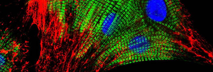
Image courtesy of Elisabeth Ehler, KCL.
Fluorescent probes or fluorophores (fluorescent dyes or proteins) are coupled to a secondary antibody or streptavidin to allow visualization of an analyte. Each fluorophore has its own spectral characteristics, with excitation and emission spectra particular to the molecule. Fluorescence facilitates the simultaneous detection of multiple analytes for a number of techniques including flow cytometry, fluorescence microscopy (epifluorescence, confocal, multiphoton and super-resolution techniques), Western blotting and ELISA.
The choice of fluorescent probe depends on a number of experimental variables, described below. There is also a Spectra Viewer Tool to assist in experimental design.
Selection of a fluorophore depends on the intended application. Sample specifics influence the choice, as they may accommodate the use of particular fluorophores, and preparation may alter the way a fluorophore behaves. The following table lists some of the considerations which are pertinent to the choice of a fluorophore for a particular technique.
| Technique | Considerations |
|---|---|
| Flow cytometry | Surface staining accommodates the use of larger and brighter fluorescent conjugates such as the fluorescent proteins (R-PE, APC and PerCP) or Brilliant Violet™ dyes. Smaller fluorescent conjugates can be used for both surface and intracellular staining. |
| IHC microscopy |
|
| IHC multiple labeling |
|
| Super-resolution microscopy (STED and STORM) |
|
| Western and dot blotting |
|
Consider excitation capabilities (lamp, lasers or LEDs determine excitation wavelengths) and the number of channels and filters available. Also consider the detection system, duration of data collection and post assay analysis.
The detection level of any fluorophore-antibody conjugate depends on brightness and photostability of the dye; antibody activity, specificity and cross-reactivity; and the optimal dye:antibody ratio (moles of dye per mole of antibody). These parameters have been researched for each of JIR’s dye conjugates to optimize the level of antibody detection and minimize background. The larger phycobiliproteins have high quantum yields (are very bright), but are limited to surface applications. Detection of poorly expressed analytes is enhanced by choosing a brighter fluorophore. For example, Alexa Fluor® 488 is brighter than FITC.
Consider autofluorescence of the sample and possible expression of recombinant fluorescently tagged proteins. Choose fluorophores whose spectra do not overlap with endogenous fluorescence.
For multiple labeling protocols, the dye panel choices will be constrained by instrumentation and sample specifics as described above. To achieve good color separation, choose fluorophores with minimal spectral overlap. The Spectra Viewer below can help build dye panels specific to any instrument.
Fluorescent probes or fluorophores (fluorescent dyes or proteins) are coupled to a secondary antibody or streptavidin to allow visualization of an analyte. Each fluorophore has its own spectral characteristics, with excitation and emission spectra particular to the molecule. Use the spectral viewer to build dye panels and compare the suitability of dyes for your application. Four dyes typically recommended for multiple labeling are pre-loaded, these can be changed as required.
For further assistance please contact our technical support team who will be delighted to assist you.
