

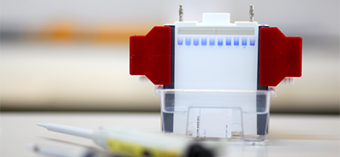
Western blotting is a technique used to confirm the presence of target proteins and peptides from complex mixtures. Proteins are separated by size using PAGE, transferred to a solid support*, and visualized using pairs of primary and secondary antibodies. Perfect your Western blots with our top 10 Western blotting tips!
(*with the exception of in-gel westerns)
- Avoid overloading your gel.
A good rule of thumb is to load only 75% of the well volume to avoid spillover into adjacent lanes. In addition, if the total protein loaded per well is excessive, the protein front may distort the migration of other protein bands, making the blot difficult to read.
- Titrate your antibodies.
The concentration of primary and secondary antibody required for each antibody pair should be optimized. Primary antibodies vary greatly. For JIR secondary antibodies, a dilution between 1:5,000 and 1:200,000 is recommended, depending on your experimental setup.
- Validate your antibodies.
Determine that your primary and secondary antibodies detect their targets under your experimental conditions using appropriate controls. A control where the primary antibody has been left out can be invaluable for confirming primary antibody-dependent signal and for background reduction.
- Block with normal serum or JIR’s high-quality (IgG- and Protease-Free) BSA.
Although skim milk may be adequate in some cases, background can be avoided by using normal serum from the host species in which your secondary antibody was produced, or JIR’s IgG- and protease-free BSA. (Learn more)
- Use a gel appropriate for the molecular weight (MW) of the proteins analyzed.
Choose a gel that will give the desired resolution between your proteins of interest. In general, 15% gels are suitable for 75 – 10 kDa, 12% for 100 – 50 kDa and 10% gels for 150 – 100 kDa proteins. Gradient gels may also be used – but remember to load appropriate protein markers!
- Dilute antibodies on the day of use.
Make up your antibody solutions on the day of use and label your tubes. We recommend centrifuging antibodies after dilution at 12,000 rpm for 30 mins.
- No AZIDE with HRP-conjugated antibodies.
Azide will inactivate the enzyme.
- Use cross-adsorbed antibodies when analyzing tissue or cell extracts.
To avoid background from cross-reactivity, choose an antibody with minimal cross-reactivity (min X) to the tissue or sample species. Find more detailed information here.
- Dilution buffers matter.
Avoid diluting your antibodies in blocking buffer, stick to diluting your antibodies in buffer only (without serum or milk). PBS containing 0.05% Tween-20 (PBS-T) is a commonly recommended dilution buffer.
- Wash adequately.
A crucial step, which may need to be optimized. In general, 3 x 10 mins in PBS-T is sufficient with a buffer volume that is suitable for the blot size.
Bonus tip
Confirm protein transfer with ponceau stain.
Stain your membranes with ponceau or similar reagents to confirm that transfer has been successful.
For more assistance with Western blot experimental design, troubleshooting, and reagent selection, see our guide or contact us.
References:
Yang, P. and Mahmood, T. (2012). Western blot: Technique, theory, and troubleshooting. North American Journal of Medical Sciences, 4(9), p.429.
Ghosh, R., Gilda, J. and Gomes, A. (2014). The necessity of and strategies for improving confidence in the accuracy of western blots. Expert Review of Proteomics, 11(5), pp.549-560.
Technical Resources | About us | Contact us | Bulk Service
Licenses | Conditions of Use | Privacy Policy | Ordering Information


FM 545248