
Rat Retina
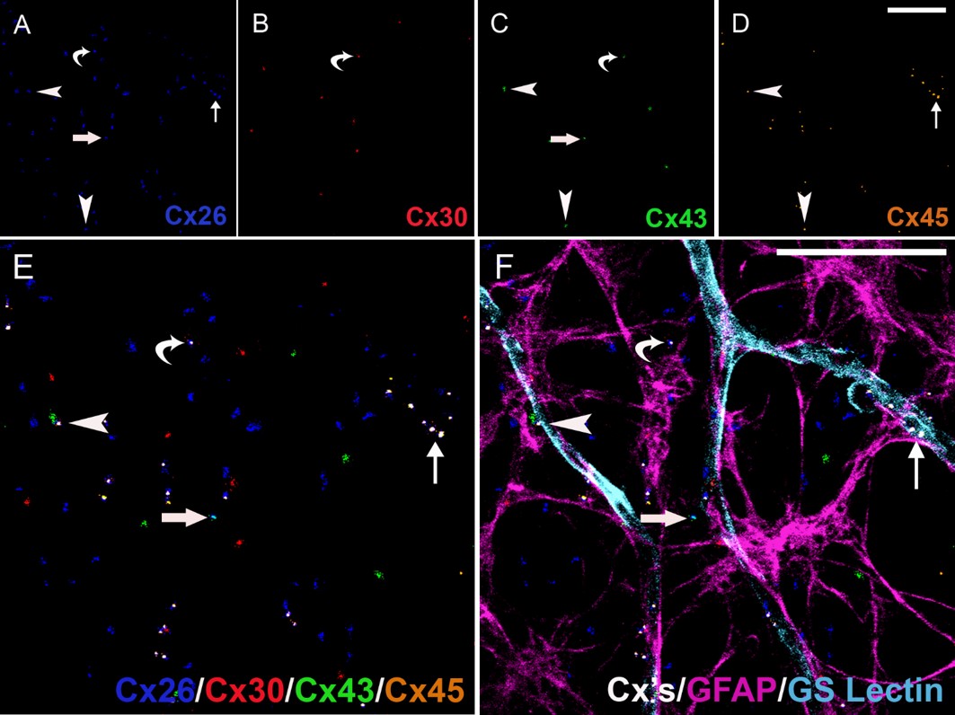
Tissue specimen of 9 month old (middle-aged) Wistar rat retina illustrates connexin expression in astrocytes in the mid-peripheral retina.
Six-marker immunohistochemistry was executed on retinal wholemount preparations. Connexin 26 (blue, Alexa Fluor® 532-conjugated anti-goat IgG), Connexin 30 (red, Cy3-conjugated anti-rabbit IgG), Connexin 43 (green, Alexa Fluor® 488-conjugated mouse IgG1), Connexin 45 (pale brown, Alexa Fluor® 594-conjugated anti-sheep IgG), GFAP stained for astrocytes (pale purple, Cy5-conjugated anti-chicken IgY), GS Isolectin B4 stained for blood vessels (turquoise, AMCA-conjugated streptavidin). Equipment used:
Carl Zeiss LSM 510 META inverted confocal microscope equipped with UV-Argon laser scanning, Axiovert 200M and LSM 510 scan head. The objective lens used was a Plan-Apochromat 63x/1.40 Oil DIC M27 and excitation laser lines (405, 488, 561 and 633 nm). Image captured of optical z-series projection, resolution of 1024 x 1024, and data depth of 12.
| Product used: | Product code: |
|---|---|
| Cy3 Donkey anti-rabbit IgG | 711-165-152 |
| Alexa Fluor® 488 goat anti-mouse IgG1 | 115-545-205 |
| Alexa Fluor® 594 Donkey anti-sheep IgG | 713-585-003 |
| Cy5 Donkey anti-chicken IgY | 703-175-155 |
| AMCA streptavidin | 016-150-084 |
References: Mansour et al (2013) Connexin 30 Expression and Frequency of Connexin Heterogeneity in Astrocyte Gap Junction Plaques Increase with Age in the Rat Retina. PLOS ONE
http://journals.plos.org/plosone/article?id=10.1371/journal.pone.0057038
Technical Resources | About us | Contact us | Bulk Service
Licenses | Conditions of Use | Privacy Policy | Ordering Information

FM 545248