
DNA-based Point Accumulation for Imaging in Nanoscale Topography (DNA-PAINT) is an increasingly popular super-resolution microscopy (SRM) technique owing to its relative simplicity. However, DNA-PAINT is hampered by slow imaging speeds and high background, which limit its utility. By modifying the original DNA-PAINT methodology to develop 2-color fluorogenic DNA-PAINT, researchers at Yale University and the University of Auckland have demonstrated a 26-fold improvement in imaging speed and increased fluorescence by as much as 57-fold, making it possible to perform 3D DNA-PAINT imaging without optical sectioning. Secondary antibodies from Jackson ImmunoResearch were used in this study.
What is DNA-PAINT?
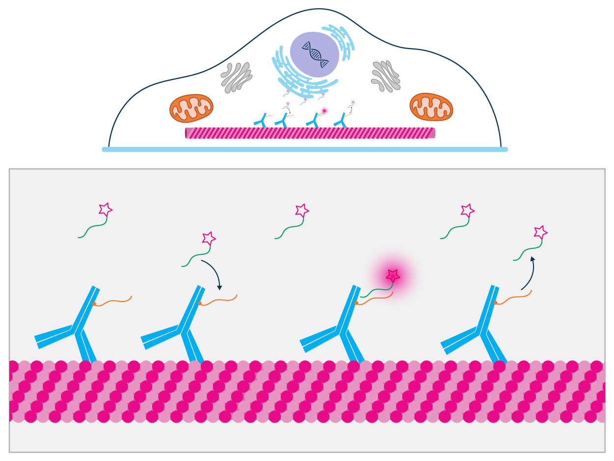
First reported by Jungmann et al. in 2010, DNA-PAINT uses transient binding events between complementary single-stranded DNA (ssDNA) oligonucleotides to localize single molecules.
One DNA strand, known as the docking strand, is linked to an antibody that binds a specific target. The other, known as the imager strand or imager probe, is covalently bound to a fluorophore and diffuses freely in solution.
When the microscope’s excitation laser is switched on, an increase in fluorescence is observed when the two strands hybridize, lasting until the imager strand unbinds. The duration of each fluorescent ‘blink’ depends on the DNA sequence, which determines the off-rate.
By capturing these transient binding events over time, it is possible to reconstruct a super-resolution image of the sample using single-molecule localization microscopy (SMLM) algorithms.
Importantly, because the fluorophores are continually replaced in the form of new imager strands, there is little risk of photobleaching.
Multiplexed imaging is achievable with the sequential addition of different pairs of docking strands/imager probes.
While the original methodology typically achieves a spatial resolution of around 10 nm, DNA-PAINT has been further developed to provide sub-nanomolar resolution.
Docking strand and imager probe design
To overcome the limitations of DNA-PAINT, Chung et al. chose to modify the design of both complementary oligonucleotides such that the imager probe would be dark when free in solution and bright when bound to the docking strand. It was hypothesized that this would not only reduce background fluorescence, but would also shorten the imaging time by allowing for an increase in imager probe concentration. As a further means of enabling rapid image acquisition, the modifications were optimized to support fast binding kinetics.
When designing the imager probe, the team exploited the fact that unbound ssDNA is highly flexible, while dsDNA takes the form of a rigid double helix. This allowed for conjugating a fluorophore (Cy3B) at the 5’ end of the imager probe and a quencher (BHQ2) at the 3’ end, resulting in strong quenching when the probe is free in solution. Upon binding to the docking strand, spatial separation of the terminals leads to a fluorescent signal.
Because this effect requires a Förster radius between the fluorophore and quencher of approximately 6 nm, the imager probe was designed to be 15 bases long. However, high off-rates for DNA-PAINT demand 10 or fewer complementary base pairs between the docking strand and the imager probe. To address this issue, Chung et al. designed a series of docking strands with internal mismatches against the imager probe, destabilizing the binding for a faster off-rate.
A second imager probe was subsequently produced, featuring ATTO 643 and the Iowa Black FQ quencher, to enable multicolor imaging. Both probes were designed to lack a self-complementary sequence, preventing stabilization in the quenched form.
Increased fluorescence
Fluorescence measurements of the probes in solution were performed using channel slides to quantify fluorescence intensity at ~10 μm deep past the coverslip. Comparison between the Cy3B/BHQ2 imager probe and a regular DNA-PAINT probe (conjugated with Cy3B but no quencher) showed the Cy3B/BHQ2 imager probe to be less than 2.5% as bright when free in solution. Importantly, the Cy3B/BHQ2 imager probe achieved a 57-fold increase in fluorescence upon binding with its fully complementary sequence, compared with just 2.4-fold for the regular probe, equating to 24 times greater fluorogenicity.
Faster imaging speeds
Imaging speed and resolution were assessed by labeling microtubules in COS-7 cells with a mouse anti-alpha tubulin primary antibody and a goat anti-mouse IgG secondary antibody (Jackson ImmunoResearch, 115-005-146), which Chung et al. conjugated to oligonucleotide docking strands using azide/DBCO click chemistry. 2D images were then captured using total internal reflection fluorescence (TIRF) microscopy. Approximately 26 times more blinking events per second could be observed with the Cy3B/BHQ2 imager probe than with the regular DNA-PAINT probe, and 2D projections of hollow microtubules were easily observed after just 3 minutes, compared with hours of imaging to obtain similar image quality with regular DNA-PAINT.
3D super-resolution without optical sectioning
When performing conventional DNA-PAINT, it is not possible to image whole cells in 3D without optical sectioning (e.g., TIRF) to suppress background fluorescence. To test whether the fluorogenic properties of the Cy3B/BHQ2 imager probe could eliminate the need for optical sectioning, Chung et al. imaged microtubules as previously described, but using regular widefield illumination and astigmatic detection for 3D localization. With the Cy3B/BHQ2 imager probe, it was possible to acquire a 3D super-resolution image of the microtubule network at high quality in just 10 minutes, with comparable localization precision to that of regular DNA-PAINT.
Simultaneous multicolor imaging
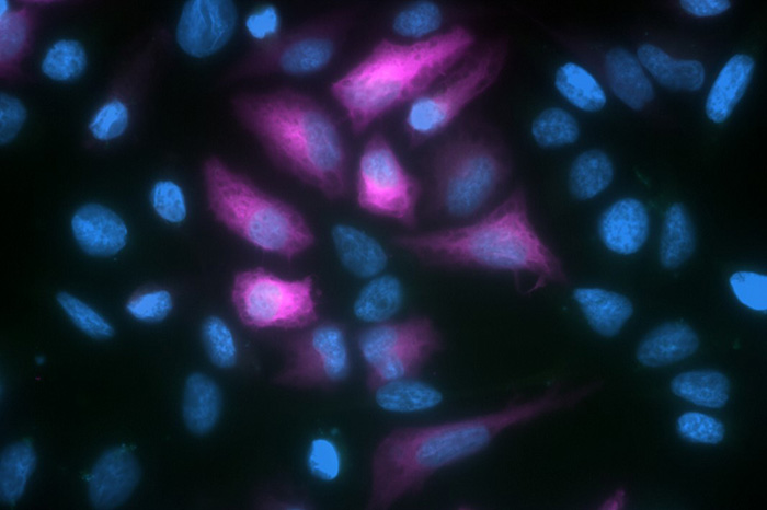
To demonstrate the utility of fluorogenic DNA-PAINT for simultaneous multicolor imaging, Chung et al. combined staining for microtubules with staining for mitochondria. Specifically, U-2 OS cells that were transfected with the GFP-labeled mitochondrial marker OMP25 were stained with a rabbit anti-GFP primary antibody and a goat anti-rabbit IgG secondary antibody (Jackson ImmunoResearch, 111-005-144) conjugated to oligonucleotide docking strands. In contrast to other multiplexed DNA-PAINT approaches that are performed sequentially, this strategy allowed for simultaneous imaging in separate color channels, reducing the time to results.
Conclusion
Fluorogenic DNA-PAINT offers several advantages over conventional DNA-PAINT, including faster imaging speeds, lower background, and the capacity for 3D super-resolution without optical sectioning. It also provides opportunities for simultaneous multicolor imaging, meaning that researchers can obtain more information from precious samples in less time.
Jackson ImmunoResearch specializes in producing secondary antibodies for life science applications, including the goat anti-mouse IgG and goat anti-rabbit IgG secondary antibodies cited in this publication. We also offer a rabbit anti-GFP antibody that can be used for DNA-PAINT and other imaging studies.

References:
- Jungmann, R., Steinhauer, C., Scheible, M., Kuzyk, A., Tinnefeld, P., & Simmel, F. C. (2010). Single-molecule kinetics and super-resolution microscopy by fluorescence imaging of transient binding on DNA origami. Nano letters, 10(11), 4756–4761. https://doi.org/10.1021/nl103427w https://pubmed.ncbi.nlm.nih.gov/20957983/
- Schnitzbauer, J., Strauss, M., Schlichthaerle, T. et al. Super-resolution microscopy with DNA-PAINT. Nat Protoc 12, 1198–1228 (2017). https://doi.org/10.1038/nprot.2017.024 https://www.nature.com/articles/nprot.2017.024
- Schueder, F., & Jungmann, R. (2024). In Situ Imaging of Proteins Using DNA-PAINT Super-Resolution Microscopy. Methods in molecular biology (Clifton, N.J.), 2800, 103–113. https://doi.org/10.1007/978-1-0716-3834-7_9 https://pubmed.ncbi.nlm.nih.gov/38709481/
