
Washing removes unbound or aggregated proteins present on the blot as well as unbound reagents that may interfere with the detection of the target molecule(s) and helps to reduce background signal. Assay sensitivity can be increased by improving the signal-to-noise ratio which can enable more accurate analysis of the protein of interest. Part 7 of our Western blotting guide details best practices for optimal washing steps for the best Western blots.
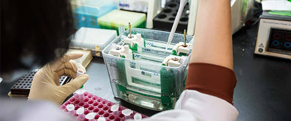
It is necessary to wash the membrane after blocking and between incubations with the antibodies. Washing removes unbound or aggregated proteins present on the blot as well as unbound reagents that may interfere with the detection of the target molecule(s). Washing helps to reduce background signal, improving assay sensitivity by increasing the signal-to-noise ratio and enabling qualitative and quantitative analysis of the protein of interest.The correct washing reagents should be used at the correct concentrations to ensure removal of deleterious agents from the membrane.
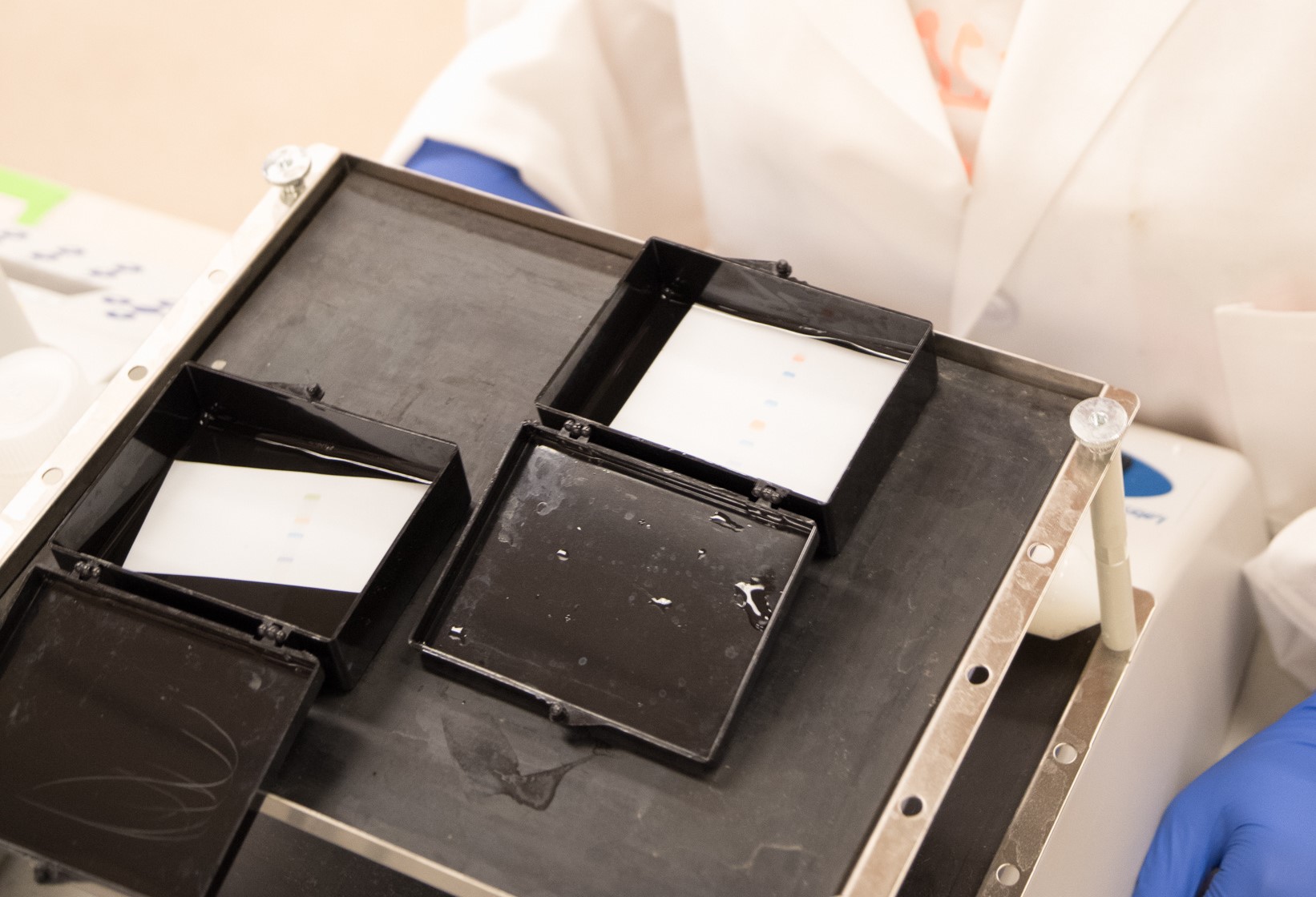
Wash Step Procedure
- Washing is usually performed at room temperature.
- The blot is washed by placing the membrane in a volume of wash buffer that fully submerges the membrane. For example, an average 10 cm² blot would need >40 ml of washing buffer, we typically use 100 ml.
- The membrane is incubated in a wash buffer for 5-10 minutes and constantly agitated by using a rocker or platform shaker or suitable blot processor.
- The washing process is then repeated between each reagent incubation.
Choosing an Appropriate Wash Buffer
Phosphate Buffered Saline (PBS)
PBS solutions can vary in their formulation, but approximate physiological conditions by combining sodium phosphate and sodium chloride to buffer the solution to a pH of 7.5. If the target protein is phosphorylated, or an Alkaline Phosphatase (AP) conjugate is to be used, PBS should be avoided as the phosphate can interact with phosphorylated proteins or quench the AP activity affecting result reliability.
Tris Buffered Saline (TBS)
TBS is sometimes used as a washing buffer as an alternative to PBS. It is typically made by combining Tris Base with Tris HCL and sodium chloride to create a solution with a physiological pH of approximately 7.2.
Detergents
Detergents such as Triton X-100, SDS, or Tween-20™ are added to prevent aggregation of unbound proteins. Sticky complexes can form and bind to the membrane, causing background Tween-20™ is typically used at 0.05% to 0.1% v/v of the solution.
Concentrations higher than 0.1% can strip the target protein(s) from the membrane and the antibodies bound to them, reducing the signal and accuracy of the blot.
Caution: Microbial Growth can create a background signal
Wash buffers should be made freshly before use.
Caution: Blocking reagents in wash buffers
Blocking reagents are sometimes added to wash buffers. We recommend using only solutions of the buffer, without the addition of blocking proteins like powdered milk or BSA.
Found Part 7 of our Western blotting guide useful? View part 8 here
References
- Blancher C. et al. (2001). SDS-PAGE and Western Blotting Techniques. Methods in Molecular Medicine. DOI: 10.1385/1-59259-136-1:145.
- Jackson ImmunoResearch Laboratories Inc. (2017). Western Blotting Troubleshooting Guide! https://www.jacksonimmuno.com/secondary-antibody-resource/technical-tips/western-blot-trouble-shooting/
- GE Healthcare. (2011). Western Blotting Principles and Methods. https://www.sigmaaldrich.com/content/dam/sigma-aldrich/docs/Sigma-Aldrich/General_Information/1/ge-western-blotting.pdf
- Bass J.J. et al. (2017). An Overview of Technical Considerations for Western Blotting Applications to Physiological Research. Scandinavian Journal of Medicine and Science in Sports. DOI: 10.1111/sms.12702
- Lewis M. (2018). An Introduction to Secondary Antibodies. Jackson ImmunoResearch Laboratories Inc. https://www.jacksonimmuno.com/secondary-antibody-resource/technical-tips/whataresecondaryantibodies/
- Lewis M. (2016). Direct and Indirect Western Blotting. Jackson ImmunoResearch Laboratories Inc. https://www.jacksonimmuno.com/secondary-antibody-resource/immuno-techniques/directandindirectwesternblotting/
- Jackson ImmunoResearch Laboratories Inc. (2020). Choosing the Right Affinity-Purified Secondary Antibody for your Application. https://www.jacksonimmuno.com/technical/products/antibody-selection
- Jackson ImmunoResearch Laboratories Inc. (2020). Multiple Labelling for Simultaneous Detection of Several Targets. https://www.jacksonimmuno.com/technical/products/protocols/multiple-labeling
- Jackson ImmunoResearch Laboratories Inc. (2020). Fluorophore Selection. https://www.jacksonimmuno.com/technical/products/conjugate-selection/fluorophore-selection

| Learn more: | Do more: |
|---|---|
| Colorimetric western blotting | Spectra Viewer |
| Chemiluminescence western blotting | Antibodies for signal enhancement |
| Fluorescent western blotting | |
Technical Resources | About us | Contact us | Bulk Service
Licenses | Conditions of Use | Privacy Policy | Ordering Information

FM 545248