Jackson Immuno Research Inc.
specializing in secondary antibodies and conjugates
Select Science Logo

Whole IgG Affinity-Purified Secondary Antibodies

"I have used a wide variety of secondaries and Jackson ImmunoResearch has consistently been the best. The fluorophores are bright and stable and their selective (x reactivity removed) secondaries have always shown species specificity in multiple labeling."

Janet Duerr, Ohio University

Rating: 5.0

Leave a Review

Alexa Fluor® 555 and 568

Contact Us

NEW Alexa Fluor® 555 and Alexa Fluor® 568 conjugates from Jackson ImmunoResearch

Flexible panel design for four-color immunostaining

Jackson ImmunoResearch now offers Alexa Fluor® 555 and 568 secondary antibody conjugates. With two new choices for the orange channel, panel design is even easier. Here, we demonstrate the utility of JIR Alexa Fluor® conjugates in two examples of four-color immunofluorescence staining protocols.

When developing an immunofluorescence experiment with multiple targets, a range of spectrally distinct fluorescent dyes and secondary antibodies that are cross-adsorbed against other species in the assay are essential for the specific detection of each target with unambiguous results.

Bright and easy to use, Alexa Fluor® dye-conjugated secondary antibodies from Jackson ImmunoResearch are now available as two new conjugates, Alexa Fluor® 555 and 568. These two new dyes complement our existing range of Alexa Fluor® conjugated secondary antibodies, offering two new choices for the orange channel.

Alongside a spectrum-wide range of fluors, Jackson ImmunoReseach Alexa Fluor® conjugated secondary antibodies are available in a broad range of target specificities. They are conjugated to Donkey or Goat host secondary antibodies that are minimally cross-reactive to many common species to avoid cross-talk between multiple primary antibodies.

Here, we demonstrate how to combine Alexa Fluor® 555 or 568 into your four-color immunofluorescence experiments.


Four-color immunofluorescence using DAPI, Alexa Fluor® 488, Alexa Fluor® 555, Alexa Fluor® 647

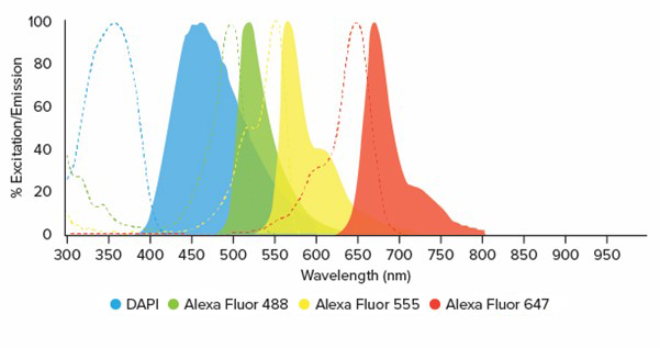
The spectral characteristics of the three Alexa Fluor® dyes in this panel (Figure 1) allow for their combination with DAPI nuclear stain for effective four-color immunofluorescence. We demonstrated this fluorophore combination by staining Human epithelial (HEp-2) cells for common intracellular protein markers. Ki-67 protein, a marker of cellular proliferation (Scholzen and Gerdes., 2000), can be seen in green. Aspects of the cytoskeleton structure can be seen in yellow and were visualized by staining microtubules using Anti-α tubulin. Cis-Golgi matrix protein, involved in the stacking of Golgi cisternae and maintenance of Golgi structure (Chang et al., 2012), was visualized using Anti Human GM130/GOLGA2 and can be seen in red at the edge of the nucleus. The nucleus was stained with DAPI and can be seen in blue. (Figure 2).

Alexa Fluor® Excitation Peak (nm) Emission Peak (nm)
DAPI 350 465
Alexa Fluor® 488 493 519
NEW! Alexa Fluor® 555 552 572
Alexa Fluor® 647 651 667
Placeholder image
Figure 1: Spectra and excitation and emission details for four-color immunofluorescence DAPI, Alexa Fluor® 488, Alexa Fluor® 555, Alexa Fluor® 647.
Placeholder image
Figure 2: Indirect four-color immunostaining of Human epithelial (HEp-2) cells. Cellular proliferation visualized in green, using Rabbit Anti-Ki-67 followed by Alexa Fluor® 488-conjugated Donkey Anti-Rabbit (H+L) (711-545-152). Microtubules visualized in yellow, using Mouse Anti-α tubulin followed by Alexa Fluor® 555 conjugated Donkey Anti-Mouse IgG (H+L) (715-565-150). Golgi structures visualized in red using Sheep Anti-Human GM130/GOLGA2, followed by Alexa Fluor® 647-conjugated Donkey Anti-Sheep IgG (H+L) (713-605-147). Blue - DAPI Nuclear Stain.

Experimental Setup:

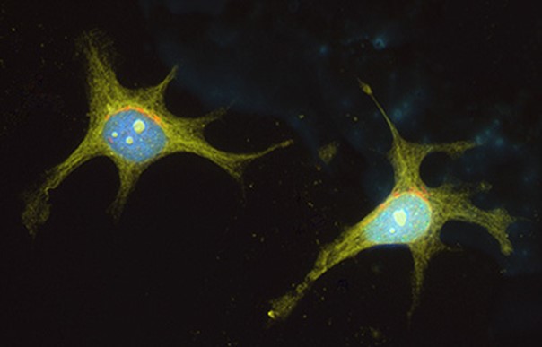
Sample: Human epithelial (HEp-2) cells , BioRad.

Primary Antibody:

  1. Rabbit Anti-Ki-67, Millipore.
  2. Mouse Anti-α-tubulin(Bov), Mol Probes.
  3. Sheep Anti-Hu GM130/GOLGA2, R&D Systems.

Secondary Antibody:

  1. Alexa Fluor® 488 Donkey Anti-Rabbit IgG (H+L) (xBov, Ck, Gt, GP, Sy Hms, Hrs, Hu, Ms, Rat, Shp), 711-545-152.
  2. Alexa Fluor® 555 Donkey Anti-Mouse IgG (H+L) (xBov, Ck, Gt, GP, Sy Hms, Hrs, Hu, Rb, Shp), 715-565-150.
  3. Alexa Fluor® 647 Donkey Anti-Sheep IgG (H+L) (xCk, GP, Sy Hms, Hrs, Hu, Ms, Rb, Rat), 713-605-147.

Nuclear Stain:

DAPI Mol Probes.

Mounting Media:

ProLong Gold antifade reagent, Invitrogen.


Four-color immunofluorescence using DAPI, Alexa Fluor® 488, Alexa Fluor® 568, Alexa Fluor® 647

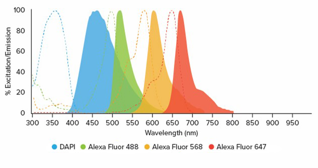
Another useful combination of dyes that can be used with DAPI for four-color immunofluorescence is Alexa Fluor® 488, 568 and 647 (Figure 3). Figure 4 demonstrates this color combination in Human epithelial (HEp-2) cells stained for common intracellular protein markers.

Ki-67 protein, a marker of cellular proliferation (Scholzen and Gerdes., 2000), can be seen in green. Aspects of the cytoskeleton structure can be seen in yellow and were visualized by staining microtubules using Anti-α tubulin. Cell-to-cell contact was visualized by staining for the epithelial cell marker, E-Cadherin protein, which can be seen in red as filaments alongside the yellow microtubules. The nucleus was stained with DAPI and can be seen in blue.

Alexa Fluor® Excitation Peak (nm) Emission Peak (nm)
DAPI 350 465
Alexa Fluor® 488 493 519
NEW! Alexa Fluor® 568 577 602
Alexa Fluor® 647 651 667
Placeholder image
Figure 3: Spectra and excitation & emission details for four-color immunofluorescence DAPI, Alexa Fluor® 488, Alexa Fluor® 568, Alexa Fluor® 647.
Placeholder image
Figure 4: Indirect four-color immunostaining of Human epithelial (HEp-2) cells. Cellular proliferation visualized in green, using Rabbit Anti-Ki-67 followed by Alexa Fluor® 488-conjugated Donkey Anti-Rabbit (H+L) (711-545-152). Microtubules visualized in yellow, using Mouse Anti-αtubulin followed by Alexa Fluor® 568 conjugated Donkey Anti-Mouse IgG (H+L) (715-575-151). Cell-to-cell contact visualized in red using Goat Anti-E-Cadherin, followed by Alexa Fluor® 647-conjugated Donkey Anti-Goat IgG (H+L) (705-605-147). Blue - DAPI Nuclear Stain.

Experimental Setup:

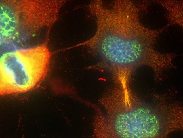
Sample: Human epithelial (HEp-2) cells, BioRad.

Primary Antibody:

  1. Rabbit A-Ki-67, Millipore.
  2. Mouse Anti-α-tubulin(Bov), Mol Probes.
  3. Goat Anti-Ms/Hu E-Cadherin, R&D Systems.

Secondary Antibody:

  1. Alexa Fluor® 488 Donkey Anti-Rabbit IgG (H+L) (xBov, Ck, Gt, GP, Sy Hms, Hrs, Hu, Ms, Rat, Shp), 711-545-152.
  2. Alexa Fluor® 568 Donkey Anti-Mouse IgG (H+L) (xBov, Ck, Gt, GP, Sy Hms, Hrs, Hu, Rb, Shp), 715-575-150.
  3. Alexa Fluor® 647 Donkey Anti-Goat IgG (H+L) (xCk, GP, Sy Hms, Hrs, Hu, Ms, Rb, Rat), 705-605-147.

Nuclear Stain:

DAPI Mol Probes.

Mounting Media:

ProLong Gold antifade reagent, Invitrogen.


Alexa Fluor® 488, 555, 568 and 647 Conjugated Antibodies


References

  • Chang, S. H., Hong, S. H., Jiang, H. L., Minai-Tehrani, A., Yu, K. N., Lee, J. H., Kim, J. E., Shin, J. Y., Kang, B., Park, S., Han, K., Chae, C., & Cho, M. H. (2012). GOLGA2/GM130, Cis-Golgi matrix protein, is a novel target of anticancer gene therapy. Molecular therapy : the journal of the American Society of Gene Therapy, 20(11), 2052–2063. https://doi.org/10.1038/mt.2012.125
  • Scholzen, T., & Gerdes, J. (2000). The Ki-67 protein: from the known and the unknown. Journal of cellular physiology, 182(3), 311–322. https://doi.org/10.1002/(SICI)1097-4652(200003)182:3<311::AID-JCP1>3.0.CO;2-9

  • Alexa Fluor® fluorescent dyes are a trademark of Life Technologies Corp. They are provided under an agreement between Life Technologies Corp and Jackson ImmunoResearch Laboratories, Inc and the manufacture, use, sale, or import of these dyes is sold pursuant to a license from Life Technologies Corp for use of its fluorescent dye technology.
go