

"I have used a wide variety of secondaries and Jackson ImmunoResearch has consistently been the best. The fluorophores are bright and stable and their selective (x reactivity removed) secondaries have always shown species specificity in multiple labeling."
Janet Duerr, Ohio UniversityRating: 5.0
Anti-IgG, Fc fragment specific antibodies may be used to detect native IgG primary antibodies without binding to the 25 kDa band of reduced and denatured IgG light chains on Western blots. Using these antibodies allows clear detection of a 25 kDa analyte, without interference from the light chains of an IP antibody. However, this detection is complicated by the appearance of degraded heavy chain antibody at 25 kDa, see Figure 1 panel A.
To avoid signal from degraded heavy chain at 25 kDa, block with monovalent Fab fragment anti-Fc (FabuLight™), see Figure 1, panels B and C. The extreme sensitivity of Western blotting requires high concentrations of the blocking reagent.
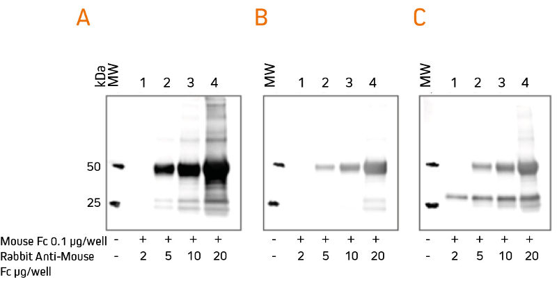
Rabbit Anti-Mouse IgG, Fcγ fragment specific (315‑005‑008) was mixed with ChromPure™ Mouse Fc (015‑000‑008) to simulate immunoprecipitation (IP).
Three identical gels were run, lanes loaded with denatured and reduced:
Lane 1: 2 μg Rabbit Anti-Mouse Fc + 0.1 μg Mouse Fc
Lane 2: 5 μg Rabbit Anti-Mouse Fc + 0.1 μg Mouse Fc
Lane 3: 10 μg Rabbit Anti-Mouse Fc + 0.1 μg Mouse Fc
Lane 4: 20 μg Rabbit Anti-Mouse Fc + 0.1 μg Mouse Fc
After SDS-PAGE and transfer to nitrocellulose, blots were blocked with BSA (10% w/v). Subsequent incubations are shown below. After incubation with secondary antibody, blots were developed with ECL substrate. Blots were imaged simultaneously, with auto exposure time based on bright bands.
No primary antibody
Secondary antibody: HRP Goat Anti-Rabbit IgG, Fc (111‑035‑008), 1:200K
Secondary antibody detects IP antibody heavy chain (HC) at 50 kDa, and degraded HC at 25 kDa.
No primary antibody
Secondary antibody: HRP Goat Anti-Rabbit IgG, Fc (111‑035‑008), 1:200K
Signal from IP antibody HC is greatly reduced. At lower loading amounts (2, 5 and 10 μg), the degraded HC at 25 kDa is not detectable.
Primary antibody: Rabbit Anti-Mouse IgG, Fcγ (min X Hu Sr Prot) (315‑035‑046), 1 μg/m
Secondary antibody: HRP Goat Anti-Rabbit IgG, Fc (111‑035‑008), 1:200K
Secondary antibody detects the primary antibody, revealing protein of interest at 25 kDa
Note that the protein of interest shows the sharpest band when amount of IP antibody is low.
Fab fragment Anti-Fc specific antibodies are found in the FabuLight tables. To block signal from a mouse IP antibody, use the Fab fragment corresponding to the subclass of the IP antibody.
