

"I have used a wide variety of secondaries and Jackson ImmunoResearch has consistently been the best. The fluorophores are bright and stable and their selective (x reactivity removed) secondaries have always shown species specificity in multiple labeling."
Janet Duerr, Ohio UniversityRating: 5.0
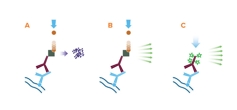
Western blotting is an analytical technique used to detect specific proteins or peptides in biological samples or solutions containing complex mixtures. Initially, linearized proteins are separated by gel electrophoresis to resolve them by size. They are then transferred onto a membrane such as nitrocellulose or polyvinylidene difluoride (PVDF) which immobilizes the protein. Subsequently, the membrane is blocked and then probed with a primary antibody directed against the specific protein of interest. A secondary antibody conjugated to a reporter molecule is then used to identify the analyte using colorimetric, chemiluminescent or fluorescent detection.
Reporter enzyme conjugates alkaline phosphatase (AP) and horseradish peroxidase (HRP) can be used for colorimetric detection. The conjugated reporter enzyme catalyzes the conversion of the chromogenic substrate to a colored precipitate, which is visualized directly on the blotting membrane. Colorimetric detection can offer quick and easily obtained results without the need for expensive detectors or extensive optimization.
Enzyme-linked conjugates can also be used for chemiluminescent signal detection. HRP conjugates produce signal by oxidizing a chemiluminescent substrate (luminol) to a form which emits light. AP conjugates produce signal when the enzyme dephosphorylates a specific substrate (e.g. 1,2-dioxetane) to a light emitting product. The signal can then be captured by exposing photographic film to the membrane or using a cooled charge-coupled device (CCD) camera. Chemiluminescent detection offers excellent sensitivity, however quantification and probing for multiple targets can be limited, and development may require refinement to optimize signal capture.
Fluorescent detection uses a fluorescent dye conjugate to visualize antigen on the membrane. Light at a wavelength specific to the dye’s spectral characteristic is absorbed, exciting the dye’s electrons to a higher electronic state, and as they return to their ground state they emit photons at the emission wavelength characteristic to the fluorophore. The light emitted is detected by a digital imager fitted with appropriate filters. Fluorescent Western blotting allows for quantitative analysis and multiplex probing without the need for stripping and reblotting. Jackson ImmunoResearch offers a range of fluorescent dyes covering the spectrum.
Jackson ImmunoResearch produces the largest diversity of species specific secondary antibody conjugates for use in Western blotting. JIR offers antibodies and streptavidin conjugated with horseradish peroxidase (HRP), alkaline phosphatase and Biotin-SP for use in traditional blots developed with chromogenic or chemiluminescent (ECL) substrates. In addition, secondary antibodies conjugated with fluorescent dyes, including far-red and infrared-emitting dyes, are available for multicolor imaging in modern readers.
Jackson ImmunoResearch also offers affinity-purified anti-horseradish peroxidase which may be used to detect HRP or to enhance signal by binding to HRP-conjugated molecules. Conjugated anti-HRP may be used to convert an HRP conjugate into a different signal.
If diluted properly, anti-light chain specific antibodies do not bind to the reduced and denatured IgG heavy chain band (50 kDa) on blots. Therefore, by using anti-light chain specific antibodies, detection of antigens with molecular weights near 50 kDa is not obscured by large amounts of reduced and denatured IgG heavy chains from primary antibodies used for immunoprecipitation (IP).
Caution: Although the antibodies react strongly with native IgG light chains, some do not react as strongly with reduced and denatured light chains on blots. Therefore, they are not recommended for sensitive and quantitative detection of reduced and denatured light chains.
If the protein of interest (POI) has a reduced and denatured molecular weight near 25 kDa, anti-IgG, Fc fragment specific antibodies may be used to detect IgG primary antibodies, without binding to reduced and denatured IgG light chains on Western blots. Clear detection of a POI near 25 kDa requires blocking degraded heavy chain that migrates to 25 kDa.
Antibodies conjugated with far-red- and infrared-emitting dyes are suitable for a variety of techniques requiring the highest sensitivity. Alexa Fluor® 680 and 790 dyes are more sensitive than visible light-emitting dyes due to lower fluorescence quenching of the conjugates and higher extinction coefficients of the dyes. Low sample autofluorescence in this region of the spectrum results in lower background signal compared with other fluorophores.
Alexa Fluor® 680 and 790 conjugates can be used for high sensitivity Western blots, quantitative Western blots, in-gel Western blots, microWestern arrays, in-cell Western assays, on-cell Western assays, and other techniques that require the brightest dyes.
Normal serum from the host species of the labeled antibody (5% v/v) is an excellent block, although 5% non-fat milk and 3% BSA are commonly used and may also be effective. Avoid milk or BSA when using a primary antibody derived from goat or sheep.
PBS/Tween 20 (0.05% v/v) or TBS/Tween, without carrier proteins, is recommended as the secondary antibody diluent. Especially when using anti-goat or anti-sheep secondary antibodies, avoid using milk or BSA in the diluent buffer. Bovine IgG in the milk or BSA may interact with the antibody due to homologous epitopes of the related species.
Recommended dilution ranges for our conjugated secondaries in Western blotting are shown in the tables below.
| Product | Chemiluminescent | Chromogenic |
|---|---|---|
| HRP-Antibody | 1:10K - 1:200K | 1:5K - 1:100K |
| Alkaline Phosphatase-Antibody | 1:5K - 1:50K | 1:5K - 1:50K |
| Biotin-Antibody | 1:20K - 1:400K | 1:20K - 1:400K |
| HRP-Streptavidin | 0.01 - 0.1 μg/mL | 1 - 2 μg/mL |
| Alkaline Phosphatase-Streptavidin | 0.1 - 1.0 μg/mL | 1 - 2 μg/mL |
| PAP | 1:5K - 1:100K | 1:5K - 1:100K |
| Product | Fluorescent |
|---|---|
| Alexa Fluor® 488, 594, 647, DyLight™ 405, and Cy™3-Antibody | 1:100 - 1:800 |
| AMCA, BV421™, BV480™, Cy2, FITC, TRITC, RRX, and Texas Red-Antibody | 1:50 - 1:200 |
| Alexa Fluor® 680 and 790-Antibody | 1:50K - 1:200K |
| Alexa Fluors®, DyLight 405, Cy3, and Cy5-Streptavidin | 0.5 - 2 µg/ml |
| Other fluorophores-Streptavidin | 2 - 5 µg/ml |
