Jackson Immuno Research Inc.
specializing in secondary antibodies and conjugates
Select Science Logo

Whole IgG Affinity-Purified Secondary Antibodies

"I have used a wide variety of secondaries and Jackson ImmunoResearch has consistently been the best. The fluorophores are bright and stable and their selective (x reactivity removed) secondaries have always shown species specificity in multiple labeling."

Janet Duerr, Ohio University

Rating: 5.0

Leave a Review

Electron Microscopy

EM with JIR Secondary Antibodies

Electron Microscopy with ImmunoGold Reagents
Figure 1. Transmission electron microscopy of fixed human cornea sections showing colocalization of MUC16 (large gold particles (12 nm Colloidal Gold Donkey Anti-Mouse IgG (H+L) (715-205-150)) and ezrin (small gold particles, arrowheads (6 nm Colloidal Gold Donkey Anti–Goat IgG (705-195-147)) on the microplicae. Scale bar, 0.1 μm. (Blalock, et al, 2007).

Electron microscopy (EM) allows the collection of high-resolution images using electron beams in the place of light. Staining and immunolabeling with electron dense material enables the visualization of the structures of the sample by adding contrast to the image. Transmission electron microscopy (TEM) requires electrons to pass through thinly sliced specimen sections, allowing 3D images to be compiled and subcellular organelles to be observed. Scanning electron microscopy (SEM) detects the scattered electrons or those emitted from the sample surface. The angle of collection gives the images a 3D quality.


Jackson ImmunoResearch ImmunoGold secondary antibodies for EM

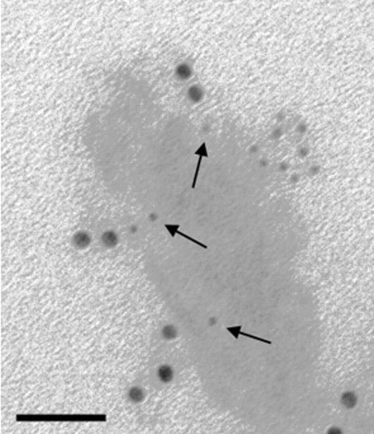
Jackson ImmunoResearch ImmunoGold reagents are colloidal gold particles complexed to secondary antibodies. The electron-dense gold increases electron scatter to reveal high contrast “spots” which allow analytes to be visualized. Labeling of multiple analytes can be achieved by using secondary antibodies complexed to differently sized particles (Figure 1). In addition to TEM and SEM applications, ImmunoGold reagents can be used for brightfield microscopy.

Read more about ImmunoGold reagents for electron microscopy and light microscopy


Localization of MUC16 in stratified HCLE cell cultures with 12 nm ImmunoGold
Figure 2. Localization of MUC16 protein on human corneal epithelial cell cultures. Localization using monoclonal Mouse Anti–Human MUC16 antibody and 12 nm Colloidal Gold Donkey Anti-Mouse IgG (H+L) (715-205-150), in stratified HCLE cell cultures. Scale bar: 0.4 μm. (Blalock, et al, 2007).

Scanning electron microscopy of human corneal epithelial cell cultures with 6 nm ImmunoGold
Figure 3. Scanning electron microscopy of the surface of human corneal epithelial cell cultures labeled with 6 nm Colloidal Gold Donkey Anti–Mouse IgG (715-195-150). (left) Non-transfected and (right) MUC16 siRNA sequence 2-transfected HCLE cells. (left, right) Field emission scanning electron microscopy showing localization of MUC16 on microplicae (insets). Scale bar: (left, right) 1 μm; (left, right insets) 0.1 μm. Images from Timothy D. Blalock, et al; Functions of MUC16 in Corneal Epithelial Cells. Invest. Ophthalmol. Vis. Sci. 2007;48(10):4509-4518.
go