"I have used a wide variety of secondaries and Jackson ImmunoResearch has consistently been the best. The fluorophores are bright and stable and their selective (x reactivity removed) secondaries have always shown species specificity in multiple labeling."
Janet Duerr, Ohio UniversityRating: 5.0
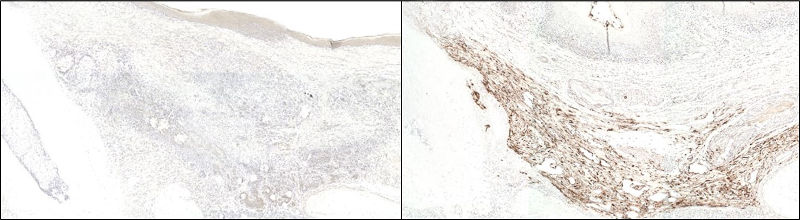
Biotin is a small molecule which non-covalently binds to avidin and streptavidin with very high affinity. The affinity of the interaction makes biotin an excellent conjugate for detection when used in immunohistochemistry techniques. Biotin conjugates can be employed in signal amplification techniques.
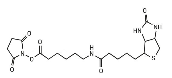
Biotin-SP is our trade name for biotin with a 6-atom spacer positioned between biotin and the protein to which it is conjugated. When Biotin-SP-conjugated antibodies are used in enzyme immunoassays, there is an increase in sensitivity compared to biotin-conjugated antibodies without the spacer. This is especially notable when Biotin-SP-conjugated antibodies are used with alkaline phosphatase-conjugated streptavidin. Apparently, the long spacer extends the biotin moiety away from the antibody surface, making it more accessible to binding sites on streptavidin.
Biotin is available conjugated to:
Biotinylated antibodies require additional reagents for visualization. We offer streptavidin and Mouse Anti-Biotin conjugated to fluorophores and enzymes.