"I have used a wide variety of secondaries and Jackson ImmunoResearch has consistently been the best. The fluorophores are bright and stable and their selective (x reactivity removed) secondaries have always shown species specificity in multiple labeling."
Imaging with AffiniPure-VHH® Fragment Alpaca Anti-GFP Antibodies
Jackson ImmunoResearch AffiniPure-VHH® Fragment Alpaca Anti-GFP antibodies are available conjugated to a range of reporter molecules, including Alexa Fluor® dyes. Designed for fluorescence microscopy, these bright dyes, combined with the polyclonal format of the AffiniPure-VHH® Anti-GFP antibodies, deliver bright target signal and spectacular signal amplification. There are many uses for AffiniPure-VHH® Fragment Alpaca Anti-GFP antibody conjugates, adding versatility to experiments or circumventing the limitations that the GFP tag can present.
Summary of the benefits of using an Anti-GFP antibody
Retrieve, rescue, and amplify the GFP signal.
Image GFP in tissue samples fixed with formalin, which can abrogate GFP fluorescence to improve sample integrity/ prevent movement of target proteins in sample.
Switch the channel to circumvent background/ endogenous GFP signal and autofluorescence.
Fluorescence flexibility - choose dyes to accommodate experimental needs and equipment.
Switching to reporter enzyme conjugate allows imaging using a non-fluorescent imaging system.
Probe for GFP in the absence of other expression/purification probes (such as His-Tag or Flag-Tag).
Enables the detection of a target protein using a universal tag when antibodies against the target protein are unavailable, unreliable, or not economically feasible.
Polyclonal Antibodies mean sensitivity and superior signal
Polyclonal detection reagents continue to offer the best sensitivity by amplifying signal, even from poorly expressing targets. Polyclonal antibodies provide a heterogeneous population of antibodies, each with a distinct paratope, thereby detecting a different portion of the GFP protein. By binding to sites across the entire protein of interest, rather than a limited specific site, region, or sequence, polyclonal AffiniPure-VHH® Anti-GFP antibodies can decorate the whole protein profusely, enabling higher labeling efficiency and a brighter signal.
Figure 1: The polyclonal format of AffiniPure-VHH® antibodies enables sensitive detection of target antigens.
Use AffiniPure-VHH® Fragment Alpaca Anti-GFP Antibodies to amplify signal
Figure 2 illustrates the utility of AffiniPure-VHH® Anti-GFP antibody conjugates. Here we demonstrate how these conjugates can be used to amplify the GFP signal and switch channels, thereby avoiding the use of a channel exhibiting autofluorescence. While Figure 3 demonstrates that even if you choose to use the GFP channel (green) with Alexa Fluor® 488 conjugated AffiniPure-VHH® Anti-GFP antibody to amplify the GFP signal, there is no discernible increase in background.
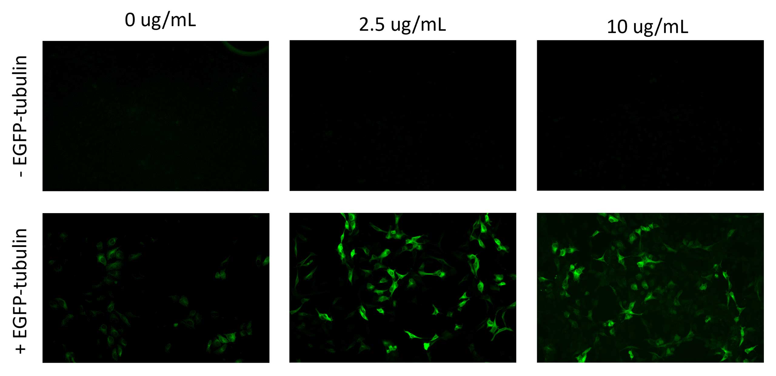
Figure 2: Immunofluorescence microscopy of mouse cortex tissue. Panel A shows wild-type mouse cortex slices. Panel B shows mouse brain cortex slices expressing a transiently transfected GFP fusion protein. Both samples were stained with 2.5 μg/mL Alexa Fluor® 647 AffiniPure-VHH® Anti-GFP antibody and DAPI. Alexa Fluor® 647 improves visualization by amplifying the GFP signal, which, without enhancement, is difficult to distinguish from the background.Figure 3: Images of HeLa cells transfected with EGFP fused to tubulin. Alexa Fluor® 488-conjugated AffiniPure-VHH® Anti-GFP antibody can enhance the signal strength, as demonstrated above. Panel A shows untransfected cells, serving as a negative control in this experiment to evaluate the background signal from the Alexa Fluor® 488 AffiniPure-VHH® Anti-GFP antibody. Panels left to right demonstrate that no increase in background is observed with increasing concentrations of the Alexa Fluor® 488 AffiniPure-VHH® Anti-GFP antibody. Images were captured with a Nikon DS-Qi2, using a 3.0x3.0 binning, an exposure time of 500 ms, and a gain of 1.0x.
Switch channels to avoid autofluorescence
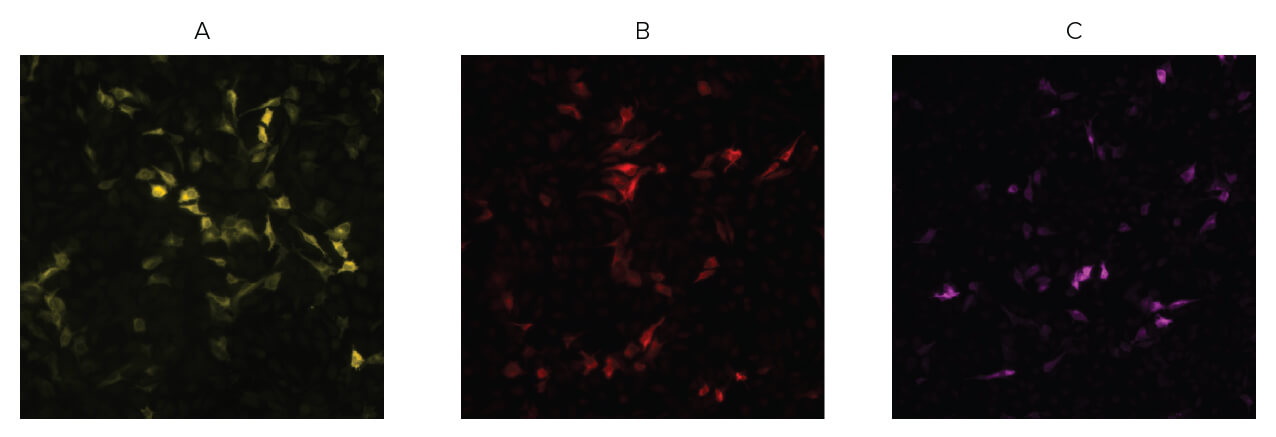
AffiniPure-VHH® Anti-GFP antibody conjugates can add versatility to an assay when working around autofluorescence or equipment limitations, such as when another target occupies a laser line or channel. Using a fluorophore that emits in a different channel, Anti-GFP antibodies can circumvent these limitations, making them useful for detecting multiple targets in the same experiment. With a wide range of conjugates available, AffiniPure-VHH® Anti-GFP antibody conjugates make it easy to switch channels and avoid autofluorescence, as orange, red, and far-red channels can be utilized (Figure 4).
Figure 4: The above shows HeLa cells transfected with EGFP-tubulin and stained with different fluorescent conjugates of AffiniPure-VHH® Anti-GFP antibody. Panel A, Alexa Fluor® 555 AffiniPure-VHH® Anti-GFP antibody (600-564-245), Panel B, Alexa Fluor® 594 AffiniPure-VHH® Anti-GFP antibody (600-584-245), Panel C, Alexa Fluor® 647 AffiniPure-VHH® Anti-GFP antibody (600-604-245). With many fluorophore options available, it is possible to avoid excitation in channels where autofluorescence occurs. All figures were captured using a Nikon JI wide-field microscope and a Fusion camera, with corresponding excitation and emission filters for each fluorophore.
Butler, Y.R., Liu, Y., Kumbhar, R. et al. α-Synuclein fibril-specific nanobody reduces prion-like α-synuclein spreading in mice. Nat Commun 13, 4060 (2022). https://doi.org/10.1038/s41467-022-31787-2
Cormack, B. P., Valdivia, R. H., & Falkow, S. (1996). FACS-optimized mutants of the green fluorescent protein (GFP). Gene, 173(1 Spec No), 33-38. https://doi.org/10.1016/0378-1119(95)00685-0
Chalfie, M., Tu, Y., Euskirchen, G., Ward, W. W., & Prasher, D. C. (1994). Green fluorescent protein as a marker for gene expression. Science (New York, N.Y.), 263(5148), 802-805. https://doi.org/10.1126/science.8303295
Kusser, K. L., & Randall, T. D. (2003). Simultaneous detection of EGFP and cell surface markers by fluorescence microscopy in lymphoid tissues. The journal of histochemistry and cytochemistry : official journal of the Histochemistry Society, 51 (1), 5-14. https://doi.org/10.1177/002215540305100102
Lippincott-Schwartz, J., Snapp, E., & Kenworthy, A. (2001). Studying protein dynamics in living cells. Nature reviews. Molecular cell biology, 2(6), 444-456. https://doi.org/10.1038/35073068
Scandella, V., Paolicelli, R. C., & Knobloch, M. (2020). A novel protocol to detect green fluorescent protein in unfixed, snap-frozen tissue. Scientific reports, 10(1), 14642. https://doi.org/10.1038/s41598-020-71493-x
SHIMOMURA, O., JOHNSON, F. H., & SAIGA, Y. (1962). Extraction, purification and properties of aequorin, a bioluminescent protein from the luminous hydromedusan, Aequorea. Journal of cellular and comparative physiology, 59, 223-239. https://doi.org/10.1002/jcp.1030590302
Snapp E. (2005). Design and use of fluorescent fusion proteins in cell biology. Current protocols in cell biology, Chapter 21, 21.4.1-21.4.13. https://doi.org/10.1002/0471143030.cb2104s27
Soboleski, M. R., Oaks, J., & Halford, W. P. (2005). Green fluorescent protein is a quantitative reporter of gene expression in individual eukaryotic cells. FASEB journal: official publication of the Federation of American Societies for Experimental Biology, 19(3), 440-442. https://doi.org/10.1096/fj.04-3180fje
Stretton, S., Techkarnjanaruk, S., McLennan, A. M., & Goodman, A. E. (1998). Use of green fluorescent protein to tag and investigate gene expression in marine bacteria. Applied and environmental microbiology, 64(7), 2554-2559. https://doi.org/10.1128/AEM.64.7.2554-2559.1998
Subramanian, S., & Srienc, F. (1996). Quantitative analysis of transient gene expression in mammalian cells using the green fluorescent protein. Journal of biotechnology, 49(1-3), 137-151. https://doi.org/10.1016/0168-1656(96)01536-2