Jackson Immuno Research Inc.
specializing in secondary antibodies and conjugates
Select Science Logo

Whole IgG Affinity-Purified Secondary Antibodies

"I have used a wide variety of secondaries and Jackson ImmunoResearch has consistently been the best. The fluorophores are bright and stable and their selective (x reactivity removed) secondaries have always shown species specificity in multiple labeling."

Janet Duerr, Ohio University

Rating: 5.0

Leave a Review

VHH Antibody Detection

Detection of VHH antibodies in common immunotechniques

Anti-VHH antibodies

Given their unique properties, VHH antibodies (nanobodies†) are growing in popularity as immunological tools. Jackson ImmunoResearch has created reagents to facilitate recognition of VHH antibodies in multiple platforms, including Western blotting, ELISA, flow cytometry, and immunofluorescence.

Read more about non-canonical immunoglobulins in camelid species.

The utility of polyclonal secondary antibodies lies in their ability to recognize multiple epitopes on a target (primary) immunoglobulin. VHH domain antibodies are obtained by immunizing goats with alpaca VHH fragments which are highly purified from naïve alpaca serum. We have shown that the antibodies have robust recognition for both alpaca and llama VHH. However, some recombinant VHH may not display many of the antigens present on native VHH fragments, making these difficult to detect with the secondary antibodies.

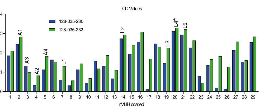
Two antibody specificities are offered. AffiniPure Goat Anti-Alpaca IgG, VHH domain (min X Bovine Serum Proteins) (128-005-232) is broadly reactive, and is recommended for screening. For multiple labeling applications, or to minimize background tissue staining, use AffiniPure Goat Anti-Alpaca IgG, VHH domain (min X Bovine, Human, Mouse, Rabbit and Rat Serum Proteins) (128-005-230). As shown in figure 3, signal strength may vary between the two versions. Fab fragments specific for the VHH domain are also available when imaging needs require smaller detection complexes.
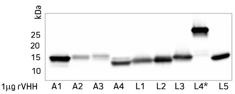
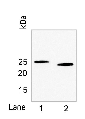
Double labeling of HEp-2 cells
Figure 1: Double labeling of HEp-2 cells for Ki-67 and Tubulin. HEp-2 cells were stained using the following combinations: Rabbit Anti-Ki-67, VHH Alpaca Anti-Rabbit (polyclonal), and Alexa Fluor® 488 conjugated Goat Anti-Alpaca IgG VHH domain (128-545-230) (green); Mouse Anti-Tubulin and RRX conj. Goat Anti- Mouse IgG (115-295-146) (red). Nuclear staining was performed using DAPI (blue).

Versatile Detection of VHH Antibodies Regardless of Species or Format

Nanobodies are commonly generated from the VHH framework of either llama or alpaca heavy chain-only antibodies. The Western blot (figure 2) shows that Goat Anti-Alpaca VHH detects both alpaca and llama VHH frameworks in their denatured state. The variation in migration reflects differences among VHH antibody structures, notably the VHH dimer L4*.

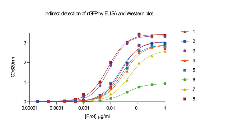
As demonstrated by ELISA (figure 3), Goat Anti-Alpaca VHH antibodies recognize the native form of both alpaca and llama rVHH. Different rVHH constructs are recognized with varying signal strength, suggesting that some rVHH do not express antigens found on the immunogen (VHH fragments from naïve alpacas). In most cases, 128-035-232 provides stronger signal than 128-035-230 because it is less highly cross-adsorbed against other species. The intended application should be considered when choosing the antibody specificity: if species cross-reactivity is not a concern, use (128-035-232) for greater sensitivity.

Western blot of different rVHH domain antibodies
Figure 2: Western blot of different rVHH domain antibodies from both alpaca and llama. Purified rVHH (1μg/well) was run by PAGE under reducing conditions and transferred to nitrocellulose. Blots were blocked with BSA (001-000-162) and probed with HRP-conjugated Goat Anti-Alpaca VHH (128-035-232) at a 1:20k dilution. ECL was used for detection.
Detection of VHH antibodies by ELISA
Figure 3: Detection of VHH antibodies from either alpaca or llama by ELISA. Each rVHH was coated onto the ELISA plate at 10 μg/ml and detected with HRP-conjugated Anti-Alpaca VHH (128-035-230 or 128-035-232) and TMB substrate.

Nanobodies are commonly generated from the VHH repertoire of either llama or alpaca heavy chain-only antibodies. The Western blot (Figure 2) shows that Goat Anti-Alpaca VHH detects both alpaca and llama VHH frameworks in their denatured state. The variation in migration reflects differences among VHH antibody structures, notably the VHH dimer L4*.

As demonstrated by ELISA (Figure 3), Goat Anti-Alpaca VHH antibodies recognize the native form of both alpaca and llama rVHH. Different rVHH constructs are recognized with varying signal strength, suggesting that some rVHH do not express epitopes found on the immunogen (VHH fragments from naïve alpacas). In most cases, 128-035-232 provides stronger signal than 128-035-230 because it is cross-adsorbed against fewer species. The intended application should be considered when choosing the antibody specificity: if species cross-reactivity is not a concern, use 128-035-232 for greater sensitivity.

VHH antibodies are growing in popularity as immunological tools. Jackson ImmunoResearch has created reagents to facilitate detection of VHH antibodies in multiple platforms, including Western blotting, ELISA, flow cytometry, and immunofluorescence.


Indirect Detection of the Protein of Interest

After a VHH antibody has bound its target, detection using a secondary antibody is an alternative to tag-directed detection. The secondary antibody provides signal amplification and the flexibility of increased conjugate options.

ELISA Detection of llama rVHH anti-GFP
Figure 4: Detection of llama rVHH anti-GFP primary antibodies by ELISA. Purified GFP was coated onto the ELISA plate at 10 μg/ml. Wells were blocked with BSA (001-000-162) and probed with serial dilutions of several rVHH anti-GFP primary antibodies. Primary antibodies were detected with HRP-conjugated Goat Anti-Alpaca VHH (128-035-232) and TMB substrate.
WB Detection of llama rVHH anti-GFP
Figure 5: Western blot showing detection of a llama rVHH anti-GFP primary antibody. Two different purified GFPs (0.5 μg/well) were run by PAGE under reducing conditions and transferred to nitrocellulose. Blots were blocked with BSA (001-000-162) and probed with rVHH anti-GFP primary antibody, followed by incubation with HRP-conjugated Goat Anti-Alpaca VHH (128-035-232) at a 1:10k dilution. ECL was used for detection.

Detection of VHH by Flow Cytometry

Recombinant VHH constructs can be candidates for therapeutic applications, creating a need to track their presence in animal models or patients. Goat Anti-Alpaca VHH antibodies (e.g. Alexa Fluor® 488 conjugate 128-545-230) can be used to detect VHH on patient cells, without recognizing endogenous IgG or primary antibodies derived from mouse, rabbit or rat. Current oncology research employs nanobody/VHH technology in a number of T cell redirection strategies to use (innate) immune defenses. These include the generation of bispecific formats to recruit and activate cytotoxic or γδ T cells, the design of nanobody-derived chimeric antigen receptor (CAR) T cells, and the development of immune checkpoint blocking nanobodies (Chanier and Chames, 2019). Characterization of the modified T-cells is typically performed by flow cytometry, allowing sorting and expansion of the cells expressing the CAR.

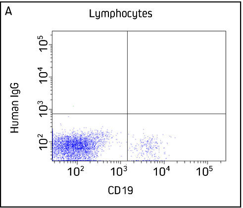
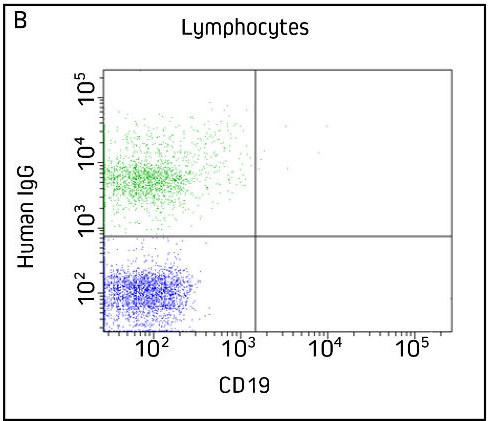
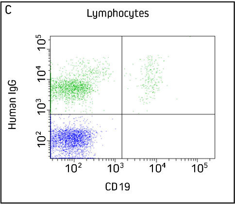
We used polyclonal alpaca VHH anti-human IgG (rather than a CAR) to demonstrate the utility of 128-545-230 in flow cytometry. The panels below represent samples gated on lymphocytes. The Goat Anti-Alpaca VHH does not recognize the mouse anti-CD19, and detects human IgG only if the VHH anti-human IgG is present.

Figure 6: Indirect detection of Human IgG on lymphocytes using Alpaca VHH Anti-Human IgG and Alexa Fluor® 488 Goat Anti-Alpaca VHH

Flow Cytometry Detection of VHH Control A
Panel A: Control, without VHH Anti-Human IgG primary antibody. Cells were incubated with Mouse anti-CD19 (APC-H7) and Alexa Fluor® 488 Goat Anti-Alpaca VHH (128-545-230) only. 128-545-230 does not recognize human IgG or the mouse primary antibody.

Flow Cytometry Detection of VHH Control B
Panel B: Control, without Mouse anti-CD19. Cells were incubated with VHH Anti-Human IgG primary antibody and Alexa Fluor® 488 Goat Anti-Alpaca VHH only. Lymphocytes displaying IgG are detected.

Flow Cytometry Detection of VHH Double Labeling
Panel C: Double labeled cells. Cells were incubated with Mouse anti-CD19 (APC-H7), VHH Anti-Human IgG primary antibody, and Alexa Fluor® 488 Goat Anti-Alpaca VHH. Lymphocytes that are positive for both human IgG and CD19 are shown in the upper right quadrant.

Imaging with VHH Antibodies

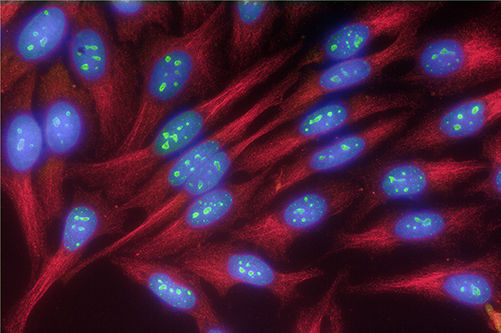
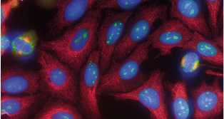
Detection using VHH antibodies requires reporter molecules such as fluorescent probes or enzymes to visualize the protein of interest. Conjugated Goat Anti-Alpaca VHH antibodies afford the inherent benefits of indirect detection such as signal amplification and conjugate choice. Goat Anti-Alpaca VHH (min X Bov, Hu, Ms, Rb, Rat Sr Prot) allows detection of VHH antibodies while minimizing tissue background and mislabeling of primary antibodies from commonly used primary antibody hosts.

For multiple labeling applications, or to minimize background tissue staining, use AffiniPure Goat Anti-Alpaca IgG, VHH domain (min X Bovine, Human, Mouse, Rabbit and Rat Serum Proteins) (128-005-230).

JIR’s 50kDa conjugated Fab fragment Anti-Alpaca IgG, VHH domain-specific antibodies can be used in combination with a primary VHH antibody to provide a binding complex much smaller than traditional antibody-antibody pairs making them favorable partners for super-resolution microscopy.

Multiple Labeling with Anti-VHH
Figure 7: Double labeling of HEp-2 cells for Ki-67 and Tubulin. HEp-2 cells were stained using the following combinations: Rabbit Anti-Ki-67, VHH Alpaca Anti-Rabbit (polyclonal), and Alexa Fluor® 488 conjugated Goat Anti-Alpaca IgG VHH domain (128-545-230) (green); Mouse Anti-Tubulin and RRX conj. Goat Anti-Mouse IgG (115-295-146) (red). Nuclear staining was performed using DAPI (blue).

Anti-VHH Secondary Antibodies

JIR’s Anti-Alpaca antibodies recognize both alpaca and llama IgG VHH domain.


References:

  • Bannas, P., Hambach, J. and Koch-Nolte, F. (2017). Nanobodies and Nanobody-Based Human Heavy Chain Antibodies As Antitumor Therapeutics. Frontiers in Immunology, 8:1603.
  • Chanier, T. and Chames, P. (2019). Nanobody Engineering: Toward Next Generation Immunotherapies and Immunoimaging of Cancer. Antibodies, 8(1):13.
  • Sircar, A., Sanni, K., Shi, J. and Gray, J. (2011). Analysis and Modeling of the Variable Region of Camelid Single-Domain Antibodies. The Journal of Immunology, 186(11), pp.6357-6367.

  • †Nanobody and nanobodies are registered trademarks of Ablynx N.V.
  • AffiniPure™ is a trademark of Jackson ImmunoResearch Laboratories, Inc.
  • DyLight™ fluorescent dyes is a trademark of Thermo Fisher Scientific.
  • Cy™ is a registered trademark of GE Healthcare.
  • Rhodamine Red™-X is a trademark of Invitrogen.
  • Alexa Fluor® is a trademark of Life Technologies Corp.
go