Find By Code Number

Product Filter

305 products available
Conjugate Selection: (Unconjugated)
Reset all
Select Science Logo

Whole IgG Affinity-Purified Secondary Antibodies

"I have used a wide variety of secondaries and Jackson ImmunoResearch has consistently been the best. The fluorophores are bright and stable and their selective (x reactivity removed) secondaries have always shown species specificity in multiple labeling."

Janet Duerr, Ohio University

Rating: 5.0

Leave a Review

AffiniPure® Rabbit Anti-GFP antibody

Contact Us

AffiniPure® Rabbit Anti-GFP antibody

Green Fluorescent Protein (GFP) is a common protein tag used to generate fusion proteins that may be expressed in prokaryotic and eukaryotic systems. The GFP tag is a useful reporter molecule because it does not require exogenous substrates or cofactors to generate fluorescence. GFP is employed in numerous applications, including quantification of gene expression, protein localization within living organisms, studying protein interactions, and as a biosensor. The addition of an Anti-GFP antibody can often improve experimental performance and overcome limitations that use of the tag itself may incur.

About Jackson ImmunoResearch AffiniPure® Rabbit Anti-GFP Antibodies

Specificity for avGFP and its derivatives

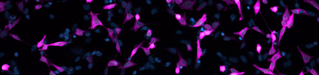
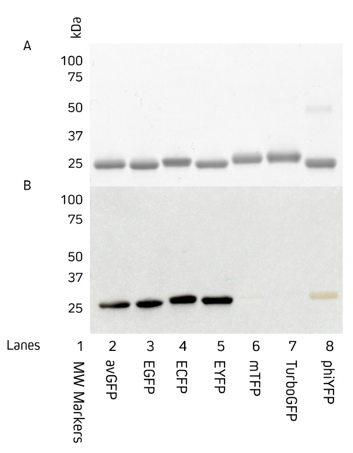
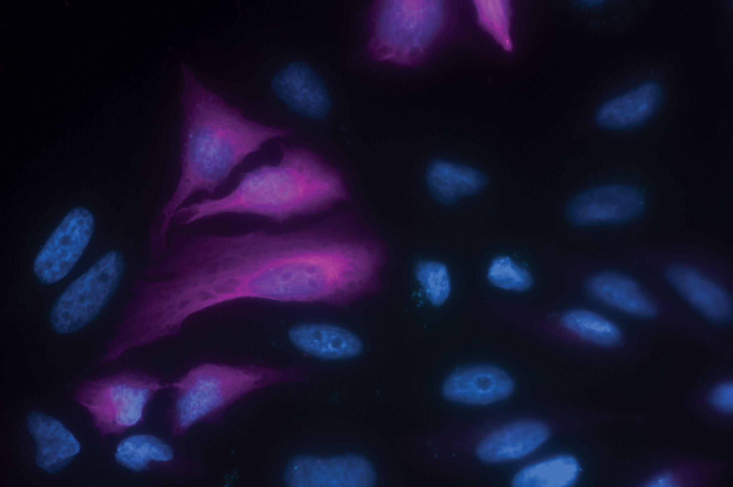
Jackson ImmunoResearch AffiniPure® Rabbit Anti-GFP is an affinity-purified polyclonal antibody raised in rabbit and can be used to detect Aequorea victoria Green Fluorescent Protein (avGFP) and its derivatives, including EGFP, ECFP, and EYFP (Fig. 1).

Figure 1: Western blot showing detection of common green fluorescent protein variants. 3 µg of purified protein was loaded onto two 12% acrylamide gels and run in tandem under reducing and denaturing conditions. The first gel, stained with Coomassie (A), shows the migration of proteins to approximately 27 kDa. The second gel (B) was used for Western Blot. This gel was transferred by electroblotting to a nitrocellulose membrane and probed with a 1:100,000 dilution of AffiniPure® HRP conjugated Rabbit Anti-GFP (300-035-245).

Versatile detection of the GFP protein across a range of applications

AffiniPure® Rabbit Anti-GFP antibody can be used to amplify the signal from GFP expressed by cells in Immunohistochemistry (IHC) or Immunofluorescence (IF) experiments. Our rabbit polyclonal Anti-GFP antibody may also be used for immunoprecipitation (IP), ELISA, ICC, Flow cytometry, and Western Blotting.

Imaging with AffiniPure® Rabbit Anti-GFP Antibodies

Conjugates for immunofluorescence

Jackson ImmunoResearch AffiniPure® Rabbit Anti-GFP antibody is available conjugated to a range of reporter molecules. For fluorescence imaging, Jackson ImmunoResearch Rabbit Anti-GFP antibodies conjugated to Alexa Fluor® dyes can deliver bright target signal and spectacular signal amplification.

Anti-GFP antibody conjugates can add versatility to an assay when trying to work around autofluorescence or equipment limitations, such as when a laser line or channel is occupied by another target. Using a fluorophore that emits in a different channel, Anti-GFP antibodies can circumvent these limitations, making them useful for detecting multiple targets in the same experiment.

Figure 2: Immunofluorescence microscopy of HeLa cells expressing a transiently transfected GFP fusion protein. Alexa Fluor® 647 Rabbit Anti-GFP detection of EGFP-tagged Tubulin protein.
Table 1: Rabbit Anti-GFP conjugates available.
Conjugate Excitation Peak (nm) Emission Peak (nm)
Alexa Fluor® 488 493 519
Alexa Fluor® 555 552 572
R-Phycoerythrin, R-PE many, 488 580
Alexa Fluor® 568 577 602
Alexa Fluor® 594 591 614
Alexa Fluor® 647 651 667
Alexa Fluor® 680 684 702
Alexa Fluor® 790 792 803
Conjugates for IHC

AffiniPure® Rabbit Anti-GFP conjugates also include popular reporter enzymes, Horseradish peroxidase (HRP), and Alkaline phosphatase, as well as a biotin conjugate facilitating the use of chromogenic IHC staining techniques and amplification protocols such as ABC (Avidin-Biotin-Complex) and LSAB (Labeled streptavidin-biotin). Switching to a non-fluorescent detection system enables the use of traditional light microscopy techniques, such as chromogenic IHC.

Product Description Product Code
Peroxidase AffiniPure® Rabbit Anti-GFP 300-035-245
Alkaline Phosphatase AffiniPure® Rabbit Anti-GFP 300-055-245
Biotin-SP (long spacer) AffiniPure® Rabbit Anti-GFP 300-065-245

Protein expression: qualification and quantification

Western Blot and ELISA (Enzyme-linked immunosorbent assays) can be used to both confirm and quantify protein expression. GFP is a popular reporter molecule used to identify successful protein expression in the absence of protein-specific antibodies. Read our article exploring the use of JIR Anti-GFP antibodies to assess protein expression qualitatively and quantitatively.

Detection of GFP by Flow Cytometry: Case study

Flow cytometry is a useful technique for screening and quantifying protein expression, particularly when they are expressed on the cell surface. The GFP tag is frequently used as a reporter molecule to track protein expression by flow cytometry. Learn how Jackson ImmunoResearch Anti-GFP antibodies can be used to detect a GFP fusion protein expressed on yeast cells by flow cytometry.


AffiniPure® Rabbit Anti-GFP Antibodies and conjugates available from Jackson ImmunoResearch

Product Description
Product Code

AffiniPure® Rabbit Anti-GFP

300-005-245

Peroxidase AffiniPure® Rabbit Anti-GFP

300-035-245

Alkaline Phosphatase AffiniPure® Rabbit Anti-GFP

300-055-245

Biotin-SP (long spacer) AffiniPure® Rabbit Anti-GFP

300-065-245

Alexa Fluor® 488 AffiniPure® Rabbit Anti-GFP

300-545-245

R-Phycoerythrin AffiniPure® Rabbit Anti-GFP

300-115-245

Alexa Fluor® 555 AffiniPure® Rabbit Anti-GFP

300-565-245

Alexa Fluor® 568 AffiniPure® Rabbit Anti-GFP

300-575-245

Alexa Fluor® 594 AffiniPure® Rabbit Anti-GFP

300-585-245

Alexa Fluor® 647 AffiniPure® Rabbit Anti-GFP

300-605-245

Alexa Fluor® 680 AffiniPure® Rabbit Anti-GFP

300-625-245

Alexa Fluor® 790 AffiniPure® Rabbit Anti-GFP

300-655-245

Table 2: AffiniPure® Rabbit Anti-GFP antibody conjugates


References

  • Cormack, B. P., Valdivia, R. H., & Falkow, S. (1996). FACS-optimized mutants of the green fluorescent protein (GFP). Gene, 173(1 Spec No), 33-38. https://doi.org/10.1016/0378-1119(95)00685-0
  • Chalfie, M., Tu, Y., Euskirchen, G., Ward, W. W., & Prasher, D. C. (1994). Green fluorescent protein as a marker for gene expression. Science (New York, N.Y.), 263(5148), 802-805. https://doi.org/10.1126/science.8303295
  • Chalfie, M. (1995), GREEN FLUORESCENT PROTEIN. Photochemistry and Photobiology, 62: 651-656. https://doi.org/10.1111/j.1751-1097.1995.tb08712.x
  • Falkow, S., Valdivia, R., Cormack, B.,(1996). FACS-optimized mutants of the green fluorescent protein (GFP). Gene,173, 1, 33-38, https://doi.org/10.1016/0378-1119(95)00685-0.
  • Kusser, K. L., & Randall, T. D. (2003). Simultaneous detection of EGFP and cell surface markers by fluorescence microscopy in lymphoid tissues. The journal of histochemistry and cytochemistry : official journal of the Histochemistry Society, 51 (1), 5-14. https://doi.org/10.1177/002215540305100102
  • Lippincott-Schwartz, J., Snapp, E., & Kenworthy, A. (2001). Studying protein dynamics in living cells. Nature reviews. Molecular cell biology, 2(6), 444-456. https://doi.org/10.1038/35073068
  • Scandella, V., Paolicelli, R. C., & Knobloch, M. (2020). A novel protocol to detect green fluorescent protein in unfixed, snap-frozen tissue. Scientific reports, 10(1), 14642. https://doi.org/10.1038/s41598-020-71493-x
  • SHIMOMURA, O., JOHNSON, F. H., & SAIGA, Y. (1962). Extraction, purification and properties of aequorin, a bioluminescent protein from the luminous hydromedusan, Aequorea. Journal of cellular and comparative physiology, 59, 223-239. https://doi.org/10.1002/jcp.1030590302
  • Snapp E. (2005). Design and use of fluorescent fusion proteins in cell biology. Current protocols in cell biology, Chapter 21, 21.4.1-21.4.13. https://doi.org/10.1002/0471143030.cb2104s27
  • Soboleski, M. R., Oaks, J., & Halford, W. P. (2005). Green fluorescent protein is a quantitative reporter of gene expression in individual eukaryotic cells. FASEB journal : official publication of the Federation of American Societies for Experimental Biology, 19(3), 440-442. https://doi.org/10.1096/fj.04-3180fje
  • Stretton, S., Techkarnjanaruk, S., McLennan, A. M., & Goodman, A. E. (1998). Use of green fluorescent protein to tag and investigate gene expression in marine bacteria. Applied and environmental microbiology, 64(7), 2554-2559. https://doi.org/10.1128/AEM.64.7.2554-2559.1998
  • Subramanian, S., & Srienc, F. (1996). Quantitative analysis of transient gene expression in mammalian cells using the green fluorescent protein. Journal of biotechnology, 49(1-3), 137-151. https://doi.org/10.1016/0168-1656(96)01536-2
  • Tsien R. Y. (1998). The green fluorescent protein. Annual review of biochemistry, 67, 509-544. https://doi.org/10.1146/annurev.biochem.67.1.509

  • AffiniPure® is a trademark of Jackson ImmunoResearch Laboratories, Inc.
  • Alexa Fluor® fluorescent dyes is a trademark of Life Technologies Corp.
go